
















©2013 Baishideng Publishing Group Co.
World J Diabetes. Dec 15, 2013; 4(6): 324-338
Published online Dec 15, 2013. doi: 10.4239/wjd.v4.i6.324
Published online Dec 15, 2013. doi: 10.4239/wjd.v4.i6.324
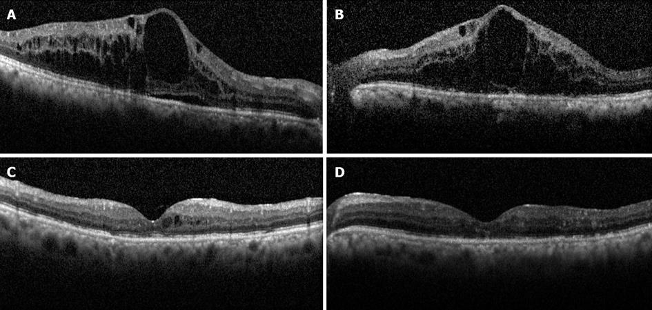
Figure 2 Horizontal spectral-domain optical coherence tomography scans of the macula before (A and B) and after (C and D) intravitreal bevacizumab therapy in a patient with diabetic macular edema.
Note extensive cystoid macular edema in both eyes (A and B) and subretinal fluid in the right (A). Six months following treatment with 3 injections of bevacizumab, the macular edema almost completely resolved in both eyes (C and D). The Snellen visual acuity improved from 20/125 to 20/70 in the right eye, but did not change significantly in the left, likely due to atrophic changes in the outer retina as seen on optical coherence tomography.
- Citation: Shamsi HNA, Masaud JS, Ghazi NG. Diabetic macular edema: New promising therapies. World J Diabetes 2013; 4(6): 324-338
- URL: https://www.wjgnet.com/1948-9358/full/v4/i6/324.htm
- DOI: https://dx.doi.org/10.4239/wjd.v4.i6.324