
















Copyright: ©Author(s) 2026.
World J Diabetes. Oct 15, 2026; 17(10): 123730
Published online Oct 15, 2026. doi: 10.4239/wjd.123730
Published online Oct 15, 2026. doi: 10.4239/wjd.123730
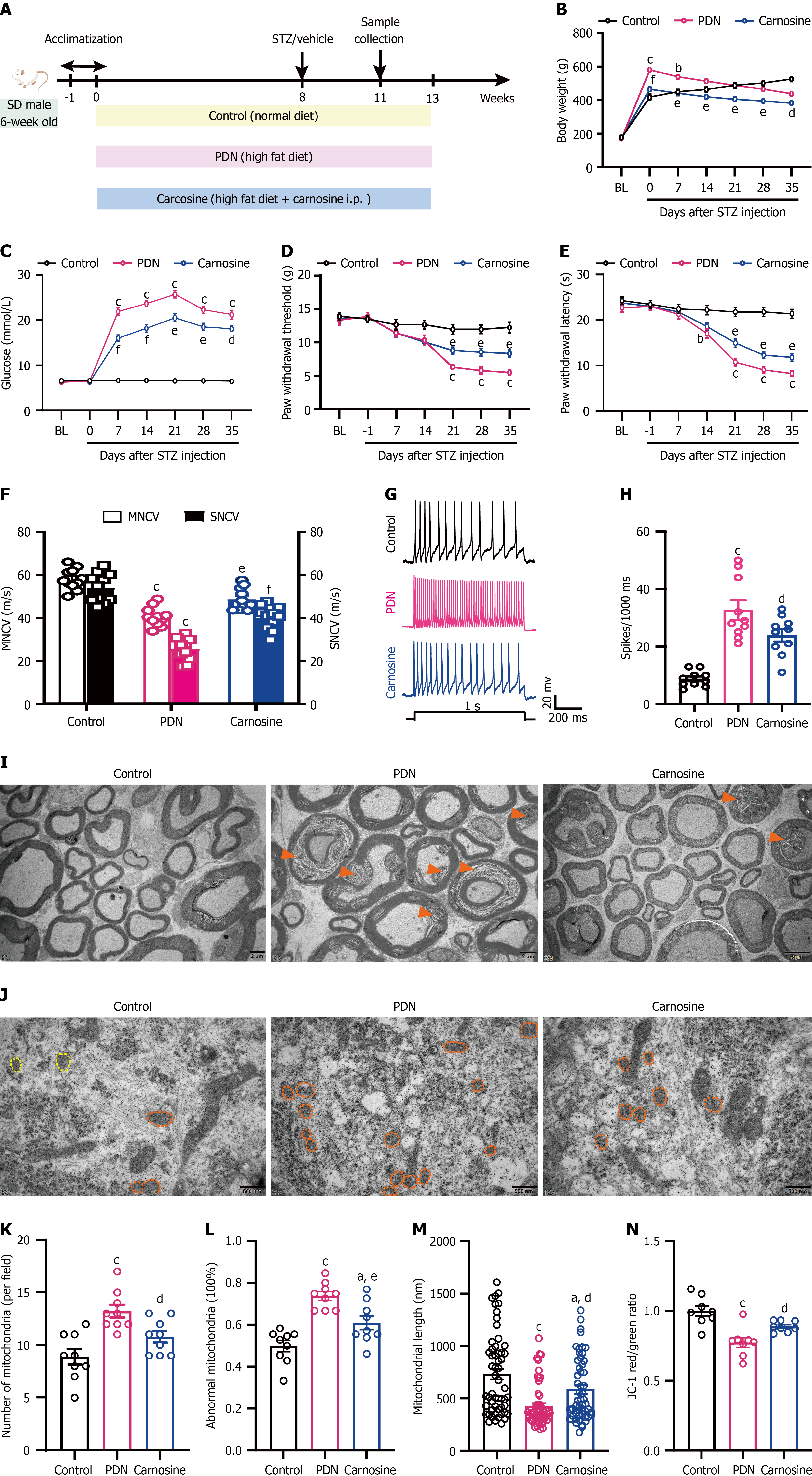
Figure 3 The impact of intraperitoneal injection of carnosine on behavioral, electrophysiological, ultrastructural, and neuronal functional abnormalities in painful diabetic neuropathy rats.
aP < 0.05, bP < 0.01, cP < 0.001 vs the control group; dP < 0.05, eP < 0.01, fP < 0.001 vs the painful diabetic neuropathy group. Two-way analysis of variance with Sidak’s multiple comparisons test for Figure 3B-E. One-way analysis of variance with Tukey’s multiple comparisons test for Figure 3F, H, and N; Linear mixed-effects models were used for statistical analyses of data shown in panels Figure 3K-M, with animal ID designated as the random intercept to account for clustered microscopic observations nested within individual rats. Bonferroni-adjusted pairwise comparisons were performed for intergroup comparisons across the three experimental groups. Figure 3B-F: n = 11; Figure 3G and H: n = 3; Figure 3I-M: n = 3 rats per group, with at least 50 mitochondria counted in 3 random fields per sample. For Figure 3K and L, each field was the statistical unit; for Figure 3M, each individual mitochondrion was the statistical unit for mitochondrial length quantification; Figure 3N: n = 8. A: Flowchart of the experiments; B: Changes in body weight after intraperitoneal carnosine injection; C: Changes in blood glucose level following intraperitoneal carnosine injection; D: Paw withdrawal threshold in rats post-intraperitoneal carnosine injection; E: Paw withdrawal latency in rats post-intraperitoneal carnosine injection; F: Motor nerve conduction velocity and sensory nerve conduction velocity in rats following intraperitoneal carnosine injection; G and H: Results of whole-cell patch-clamp recordings at week 3 after streptozotocin injection; G: Representative traces of neuronal action potential; H: Statistical analysis of the action potential number in dorsal root ganglia (DRG) neurons of rats (n = 3); I: Ultrastructural changes of the DRG post-intraperitoneal carnosine injection. Scale bar = 2 μm; J: Alterations in mitochondrial structure in the DRG post-intraperitoneal carnosine injection. Scale bar = 500 nm; K: The number of mitochondria per field; L: Proportion of abnormal mitochondria relative to the total mitochondrial count; M: Mitochondrial length; N: Changes in mitochondrial membrane potential in the DRG following intraperitoneal carnosine injection. PDN: Painful diabetic neuropathy; MNCV: Motor nerve conduction velocity; SNCV: Sensory nerve conduction velocity; STZ: Streptozotocin.
- Citation: Yu ZY, Yang J, Jiang Y, Han YZ, Wei M, Shou JY, Shen SX, Lyu YH, Li XS, Chen Q, Zhao XR, Xing GG, Ma DQ, Li M. Preventive carnosine supplementation attenuates painful diabetic neuropathy via restoring mitochondrial function and suppressing inflammasome activation. World J Diabetes 2026; 17(10): 123730
- URL: https://www.wjgnet.com/1948-9358/full/v17/i10/123730.htm
- DOI: https://dx.doi.org/10.4239/wjd.123730