
















©The Author(s) 2026.
World J Diabetes. Jan 15, 2026; 17(1): 112942
Published online Jan 15, 2026. doi: 10.4239/wjd.v17.i1.112942
Published online Jan 15, 2026. doi: 10.4239/wjd.v17.i1.112942
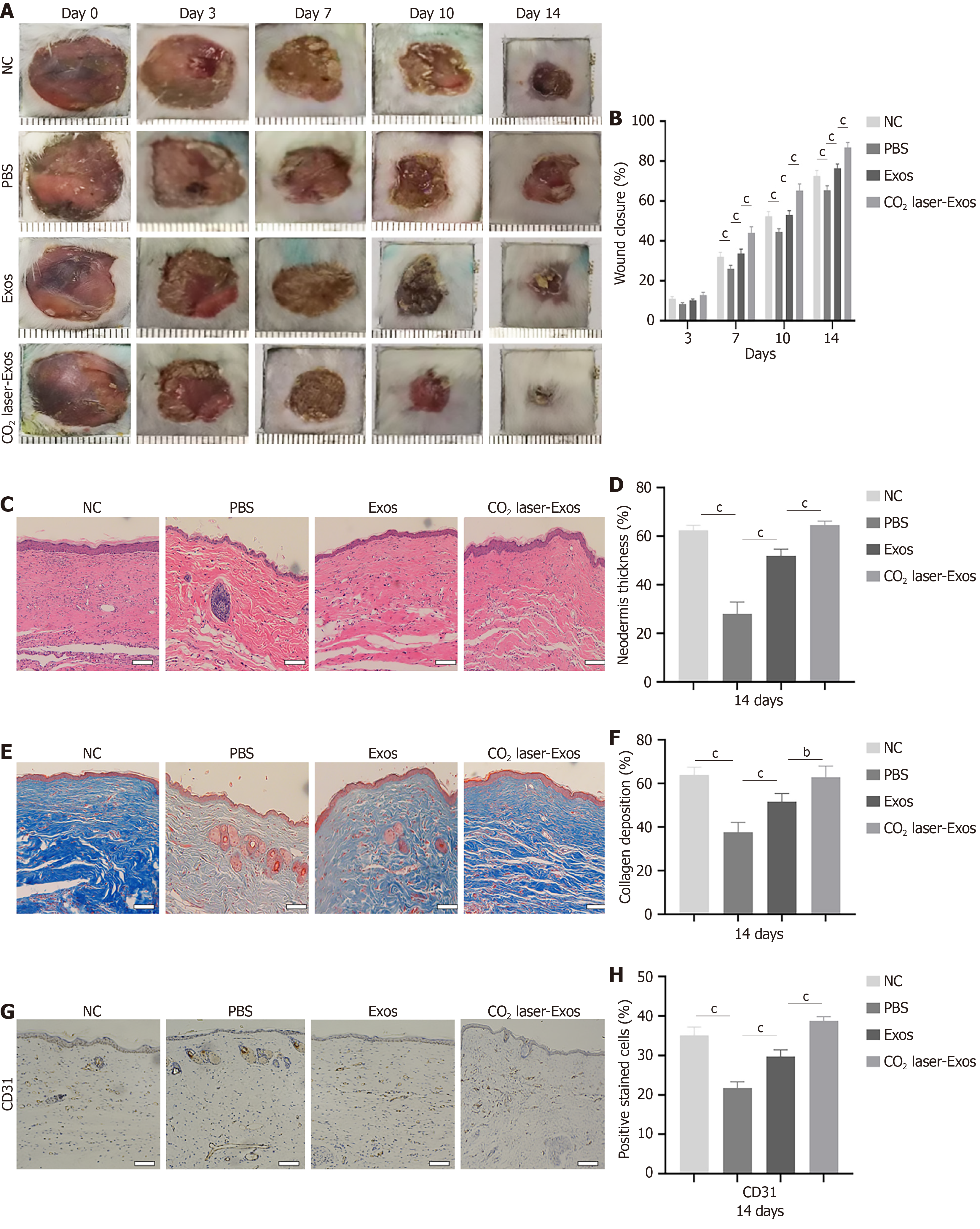
Figure 8 Exosomes derived from CO2 laser-preconditioned adipose-derived mesenchymal stem cells accelerate angiogenesis in diabetic wounds.
bP < 0.01; cP < 0.001. Data are presented as mean ± SD (n = 6). A and B: Representative images of full-thickness skin defects in non-diabetic mice and diabetic mice treated with phosphate-buffered saline, exosomes, or exosomes derived from CO2 laser-preconditioned adipose-derived mesenchymal stem cells on 0 day, 3 days, 7 days, 10 days, and 14 days post-surgery; C and D: Quantification of hematoxylin and eosin staining and skin thickness on day 14. Scale bar = 100 μm; E and F: Quantification of Masson’s trichrome staining and collagen deposition on day 14. Scale bar = 100 μm; G and H: Representative immu
- Citation: Chen JY, Ji Z, Guo K, Wang HN, Zhu CC, Li T, Zhao XB, Wang YT, Li Q, Jin PS, Li XY. Fractional carbon dioxide laser-induced photothermal activation of mesenchymal stem cell-derived exosomes accelerates diabetic wound healing by enhancing angiogenesis. World J Diabetes 2026; 17(1): 112942
- URL: https://www.wjgnet.com/1948-9358/full/v17/i1/112942.htm
- DOI: https://dx.doi.org/10.4239/wjd.v17.i1.112942