
















©The Author(s) 2026.
World J Diabetes. Jan 15, 2026; 17(1): 112942
Published online Jan 15, 2026. doi: 10.4239/wjd.v17.i1.112942
Published online Jan 15, 2026. doi: 10.4239/wjd.v17.i1.112942
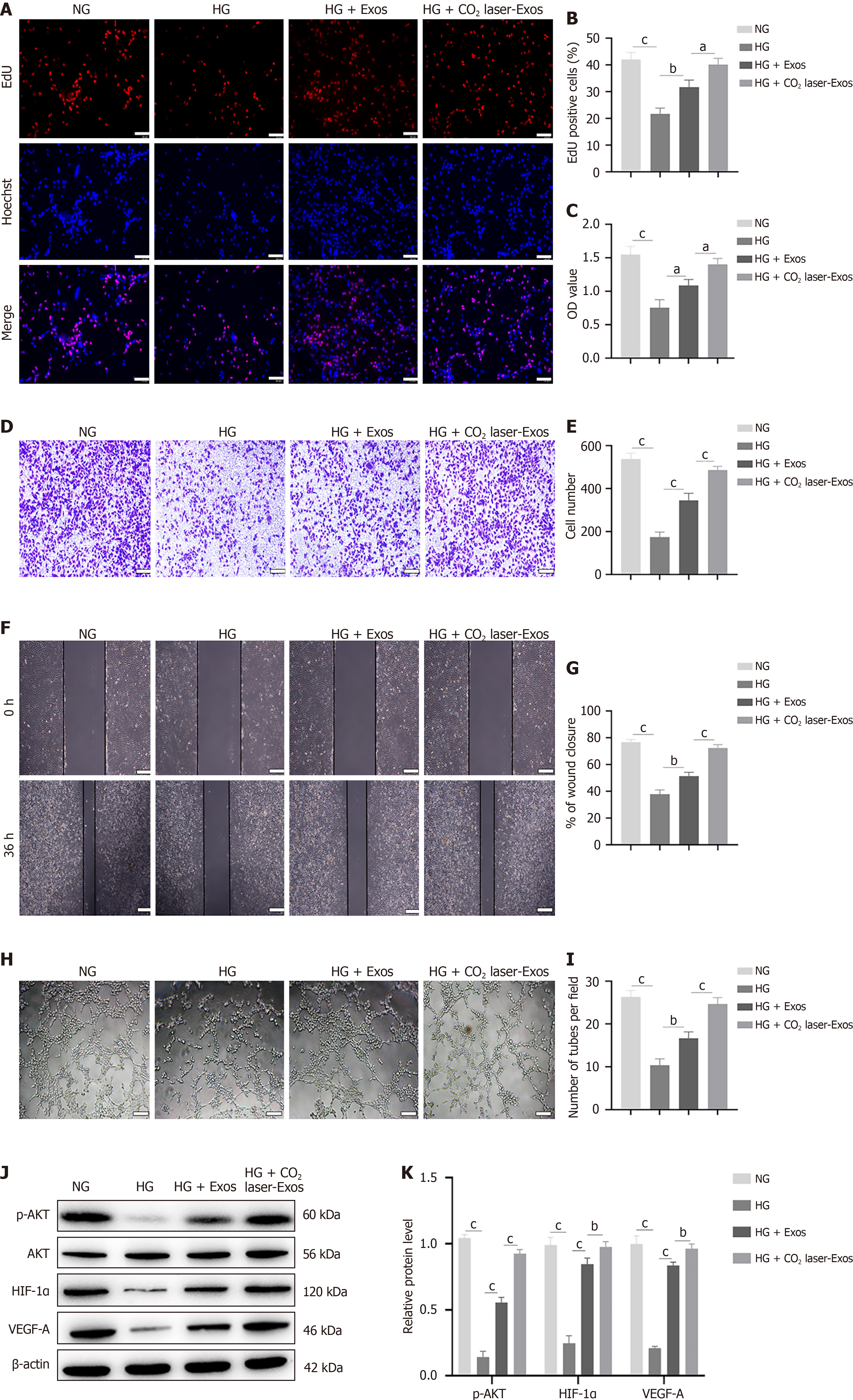
Figure 4 CO2 laser-exosomes promote angiogenesis in human umbilical vein endothelial cells.
aP < 0.05, bP < 0.01, cP < 0.001. Data are presented as mean ± SD (n = 3). A and B: 5-ethynyl-2’-deoxyuridine incorporation assay to assess the proliferative capacity of human umbilical vein endothelial cells (HUVECs). Scale bar = 100 μm; C: Cell Counting Kit-8 assay to evaluate the viability of HUVECs after treatment with high glucose and exosomes; D and E: Transwell migration assay to evaluate the migratory ability of HUVECs. Scale bar = 200 μm; F and G: In vitro wound healing assay to assess cell migration. Scale bar = 200 μm; H and I: Tube formation assay to evaluate the ability of HUVECs to form capillary-like structures. Scale bar = 200 μm; J and K: Western blot analysis of the expression levels of phosphorylated protein kinase B, protein kinase B, hypoxia-inducible factor-1α, and vascular endothelial growth factor-A. NG: Normal glucose; HG: High glucose; Exos: Exosomes; CO2 laser-Exos: Exosomes derived from CO2 laser-preconditioned adipose-derived mesenchymal stem cells; EdU: 5-Ethynyl-2’-deoxyuridine; AKT: Protein kinase B; p-AKT: Phosphorylated protein kinase B; HIF-1α: Hypoxia-inducible factor-1α; VEGF-A: Vascular endothelial growth factor-A.
- Citation: Chen JY, Ji Z, Guo K, Wang HN, Zhu CC, Li T, Zhao XB, Wang YT, Li Q, Jin PS, Li XY. Fractional carbon dioxide laser-induced photothermal activation of mesenchymal stem cell-derived exosomes accelerates diabetic wound healing by enhancing angiogenesis. World J Diabetes 2026; 17(1): 112942
- URL: https://www.wjgnet.com/1948-9358/full/v17/i1/112942.htm
- DOI: https://dx.doi.org/10.4239/wjd.v17.i1.112942