Copyright
©The Author(s) 2023.
World J Diabetes. Aug 15, 2023; 14(8): 1323-1329
Published online Aug 15, 2023. doi: 10.4239/wjd.v14.i8.1323
Published online Aug 15, 2023. doi: 10.4239/wjd.v14.i8.1323
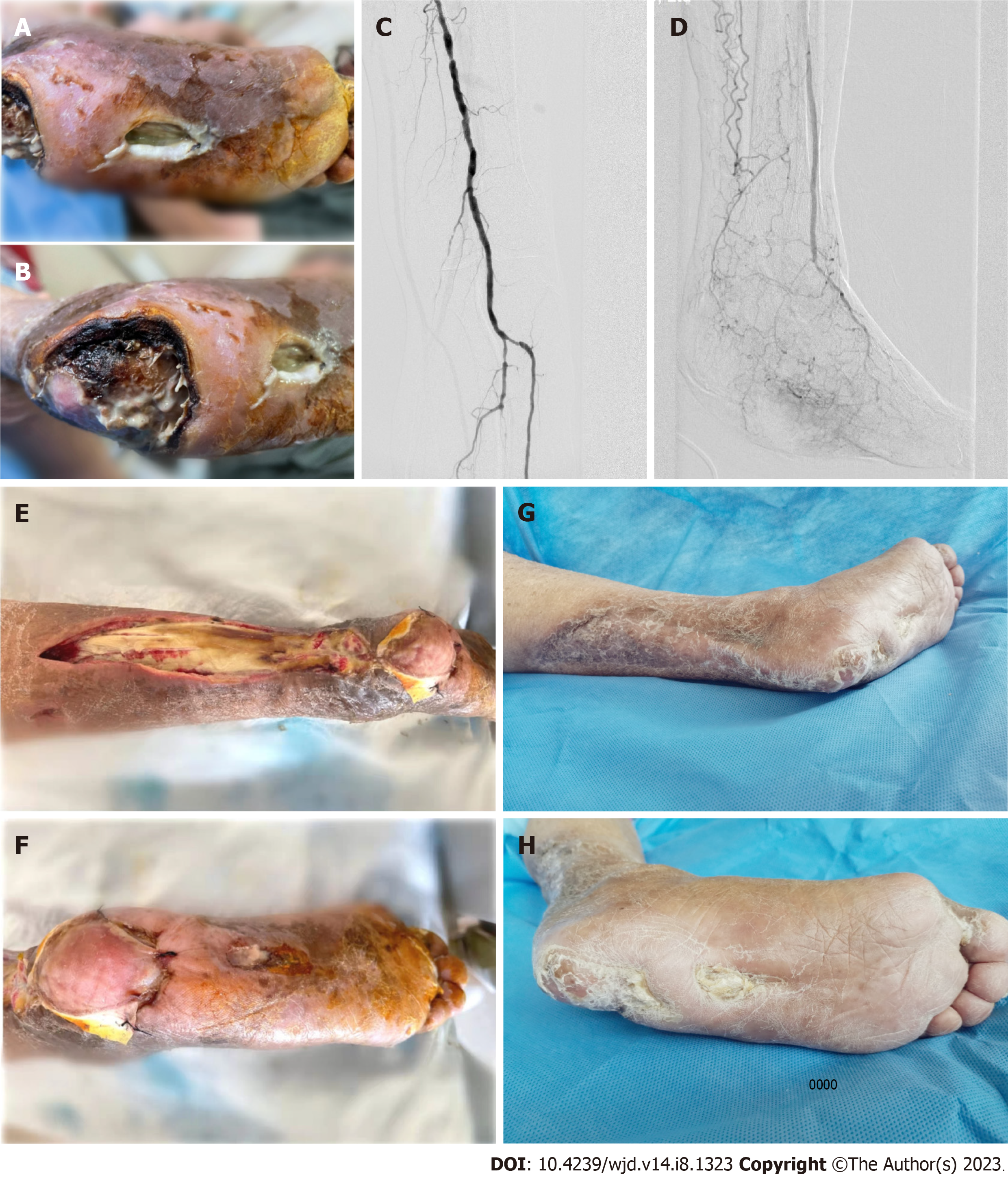
Figure 1 Management of lower limb ulcer and arterial stenosis.
A: Wound in the middle of the planta pedis, with plantar fascia exposed; B: Heel ulcer, which is connected to the wound in the middle of the planta pedis; C: Extensive arterial stenosis and occlusion in the left lower limb shown by digital subtraction angiography; D: Blood flow of the foot after percutaneous transluminal angioplasty; E: The ulcer spreading to the lower leg, and the wound reconstruction on the back of the lower leg; F: Application of tissue-engineered skin to the heel wound; G: Wound healing on the back of the lower leg; H: Wound healing on the planta pedis.
- Citation: Wang JJ, Yu YY, Wang PY, Huang XM, Chen X, Chen XG. Sequential treatment for diabetic foot ulcers in dialysis patients: A case report. World J Diabetes 2023; 14(8): 1323-1329
- URL: https://www.wjgnet.com/1948-9358/full/v14/i8/1323.htm
- DOI: https://dx.doi.org/10.4239/wjd.v14.i8.1323


