Copyright
©The Author(s) 2017.
World J Diabetes. May 15, 2017; 8(5): 187-201
Published online May 15, 2017. doi: 10.4239/wjd.v8.i5.187
Published online May 15, 2017. doi: 10.4239/wjd.v8.i5.187
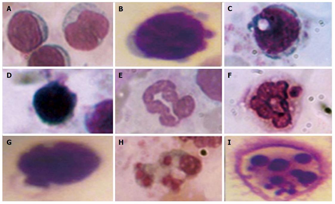
Figure 1 Morphological features of leukocytes apoptosis.
Lymphocytes: Normal cell without apoptotic sings (А), zeiosis of the membrane (B), vacuolization of the cytoplasm (C), karyopyknozis of the nucleus (D). Neutrophils: Normal cell without apoptotic sings (E), vacuolization of the cytoplasm and the nucleus (F, G), vacuolization of the cytoplasm (H) and karioreksis of the nucleus (I). In smears stained by the Romanovsky-Himza method, the number of white blood cells with features of apoptosis was assessed. The ratio between cells with morphological apoptotic features and the general quantity of cells were expressed in percentages.
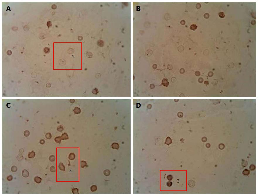
Figure 2 Immunocytochemical analysis of peripheral blood leukocytes in rats depending on the content of p53 pro-apoptotic protein in healthy animals, animals with streptozotocin-induced diabetes and treated with submerged cultured mycelium powder of mushrooms.
A: Control; B: Control animals treated with Agaricus brasiliensis (A. brasiliensis); С: Animals with STZ-induced diabetes mellitus; D: Diabetic animals treated with A. brasiliensis. 1: p53-; 2: p53+; 3: p53++. An indirect immunoperoxidase method was used for detection and visualization of intracellular protein p53. The content analysis of p53 in leukocytes of rat peripheral blood was performed by light microscopy using a × 40 microscope objective. Depending on intensity of staining, the cells were divided into 3 groups: Negative reaction (p53-), positive reaction (p53+), and high-positive (p53++) reaction.
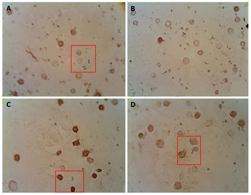
Figure 3 Immunocytochemical analysis of peripheral blood leukocytes in rats depending on the content of anti-apoptotic Bcl-2 protein in healthy animals, animals with streptozotocin-induced diabetes and treated with the submerged cultured mycelium powder of mushrooms.
A: Control; B: Control animals treated with G. lucidum; С: Animals with STZ-induced DM; D: Diabetic animals treated with G. lucidum. 1: Bcl-2-; 2: Bcl-2+; 3: Bcl-2++. An indirect immunoperoxidase method was used for detection and visualization of intracellular proteins Bcl-2. The content analysis of Bcl-2 in leukocytes of rat peripheral blood was performed by light microscopy using a × 40 microscope objective. Depending on intensity of staining, the cells were divided into 3 groups: Negative reaction (Bcl-2-), positive reaction (Bcl-2+), and high-positive (Bcl-2++) reaction. G. lucidum: Ganoderma lucidum; STZ: Streptozotocin; DM: Diabetes mellitus.
- Citation: Vitak T, Yurkiv B, Wasser S, Nevo E, Sybirna N. Effect of medicinal mushrooms on blood cells under conditions of diabetes mellitus. World J Diabetes 2017; 8(5): 187-201
- URL: https://www.wjgnet.com/1948-9358/full/v8/i5/187.htm
- DOI: https://dx.doi.org/10.4239/wjd.v8.i5.187


