Copyright
©The Author(s) 2017.
World J Gastrointest Oncol. Sep 15, 2017; 9(9): 390-396
Published online Sep 15, 2017. doi: 10.4251/wjgo.v9.i9.390
Published online Sep 15, 2017. doi: 10.4251/wjgo.v9.i9.390
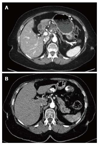
Figure 1 Computed tomography image.
A: Computed tomography (CT) image of the solid-cystic pancreatic mass with distal atrophy of the pancreas and pancreatic duct dilatation; B: CT three years before in which no pancreatic lesions were present.
- Citation: Martínez de Juan F, Reolid Escribano M, Martínez Lapiedra C, Maia de Alcantara F, Caballero Soto M, Calatrava Fons A, Machado I. Pancreatic adenosquamous carcinoma and intraductal papillary mucinous neoplasm in a CDKN2A germline mutation carrier. World J Gastrointest Oncol 2017; 9(9): 390-396
- URL: https://www.wjgnet.com/1948-5204/full/v9/i9/390.htm
- DOI: https://dx.doi.org/10.4251/wjgo.v9.i9.390


