Copyright
©The Author(s) 2017.
World J Gastrointest Oncol. Nov 15, 2017; 9(11): 444-451
Published online Nov 15, 2017. doi: 10.4251/wjgo.v9.i11.444
Published online Nov 15, 2017. doi: 10.4251/wjgo.v9.i11.444
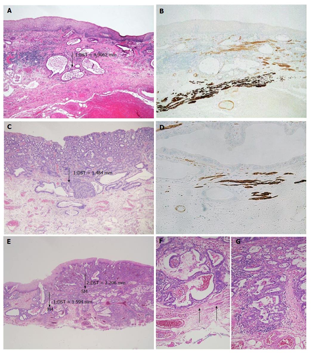
Figure 2 In case of undermining growth beneath the intact epithelium, tumour thickness was measured from the most superficial neoplastic cell layer to the point of deepest invasion.
A: HE 50 ×; ESD specimen with early Barrett’s adenocarcinoma pT1 (m3) with infiltration between the SMM and DMM and a tumour thickness of 900 μm. Tumour thickness was measured from the most superficial tumour cell layer to the deepest point of the invasion; B: Smoothelin IHC 50 ×; Immunohistochemical staining discriminates between the SMM (light brown) and DMM (dark brown). A tumour gland can be seen in-between the two muscle layers; C: HE 16 ×; Neoplastic glands reach the DMM. Smooth muscle fibres are found in the neighbourhood of the glands. The tumour thickness is approximately 1500 μm (m4); D: Smoothelin IHC 200 ×; Immunohistochemical staining confirms the m4 stage. Dark brown fibres of the DMM are found on the same level as the tumour glands; E: HE 16 ×; ESD specimen with an adenocarcinoma of the oesophagus that reaches the submucosa. The left-sided measurement was performed in an area where the tumour was restricted to a m4-stage (tumour thickness approxinately 1600 μm). The right-sided measurement was in an area where the tumour already showed the beginning of an infiltration of the submucosa (tumour thickness approxinately 2200 μm); F: HE 100 ×; Higher magnification of the m4-area of (E). Smooth muscle fibres (arrows) discriminate from sm-stage; G: HE 100 ×; Higher magnification of the sm-area of (E). The lack of muscle fibres indicates the sm-stage. ESD: Endoscopic submucosa dissection; SMM: Superficial muscularis mucosae; DMM: Deep muscularis mucosae; IHC: Immunohistochemistry; HE: Haematoxylin and eosin.
- Citation: Endhardt K, Märkl B, Probst A, Schaller T, Aust D. Value of histomorphometric tumour thickness and smoothelin for conventional m-classification in early oesophageal adenocarcinoma. World J Gastrointest Oncol 2017; 9(11): 444-451
- URL: https://www.wjgnet.com/1948-5204/full/v9/i11/444.htm
- DOI: https://dx.doi.org/10.4251/wjgo.v9.i11.444


