
















©2014 Baishideng Publishing Group Inc.
World J Gastrointest Oncol. Sep 15, 2014; 6(9): 330-343
Published online Sep 15, 2014. doi: 10.4251/wjgo.v6.i9.330
Published online Sep 15, 2014. doi: 10.4251/wjgo.v6.i9.330
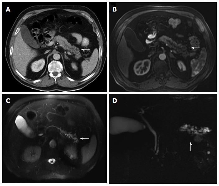
Figure 20 Multidetector computed tomography image.
A: Cystic dilatation of the main pancreatic duct and some of its branches in the pancreatic tail. Ductal communication with the tumor cannot be clearly identified; B-D: In contrast-enhanced axial T1 (B) and T2-weighted (C) magnetic resonance images and in magnetic resonance imaging cholangiography (D) ductal communication can be easily detectable.
- Citation: Santa LGL, Retortillo JAP, Miguel AC, Klein LM. Radiology of pancreatic neoplasms: An update. World J Gastrointest Oncol 2014; 6(9): 330-343
- URL: https://www.wjgnet.com/1948-5204/full/v6/i9/330.htm
- DOI: https://dx.doi.org/10.4251/wjgo.v6.i9.330