Copyright
©2014 Baishideng Publishing Group Inc.
World J Gastrointest Oncol. Aug 15, 2014; 6(8): 301-310
Published online Aug 15, 2014. doi: 10.4251/wjgo.v6.i8.301
Published online Aug 15, 2014. doi: 10.4251/wjgo.v6.i8.301
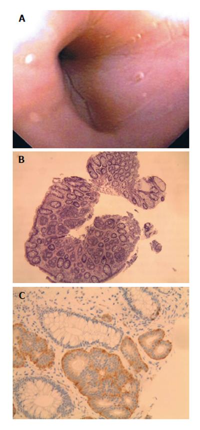
Figure 4 A 50-year-old female presented for evaluation of gastroesophageal reflux disease and colon cancer screening.
A: Patient 7, neuroendocrine (carcinoid) tumors as sessile sigmoid polyp at colonoscopy; B: Organoid growth pattern with regular bland nuclei with indistinct cell borders. H and E, ×10; C: The neuroendocrine cells are positive for Synaptophysin and adjacent colonic glands are negative. Synaptophysin, × 20.
- Citation: Salyers WJ, Vega KJ, Munoz JC, Trotman BW, Tanev SS. Neuroendocrine tumors of the gastrointestinal tract: Case reports and literature review. World J Gastrointest Oncol 2014; 6(8): 301-310
- URL: https://www.wjgnet.com/1948-5204/full/v6/i8/301.htm
- DOI: https://dx.doi.org/10.4251/wjgo.v6.i8.301


