Copyright
©2014 Baishideng Publishing Group Inc.
World J Gastrointest Oncol. Jul 15, 2014; 6(7): 225-243
Published online Jul 15, 2014. doi: 10.4251/wjgo.v6.i7.225
Published online Jul 15, 2014. doi: 10.4251/wjgo.v6.i7.225
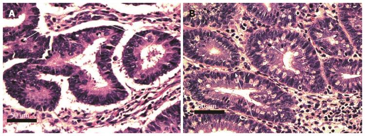
Figure 7 Crypts of tubular adenomas with high grade dysplasia cut across the short axis, human (A) and mouse (B).
Glands with high grade dysplasia show overlapping cells with oval to round vesicular nuclei and prominent nucleoli (long arrows). Mitotic figures are abundant (short arrows). Complex architecture with infolding of crypts can also be seen. Images obtained with 40× objective lens.
- Citation: Prasad AR, Prasad S, Nguyen H, Facista A, Lewis C, Zaitlin B, Bernstein H, Bernstein C. Novel diet-related mouse model of colon cancer parallels human colon cancer. World J Gastrointest Oncol 2014; 6(7): 225-243
- URL: https://www.wjgnet.com/1948-5204/full/v6/i7/225.htm
- DOI: https://dx.doi.org/10.4251/wjgo.v6.i7.225


