Copyright
©2010 Baishideng Publishing Group Co.
World J Gastrointest Oncol. Sep 15, 2010; 2(9): 364-368
Published online Sep 15, 2010. doi: 10.4251/wjgo.v2.i9.364
Published online Sep 15, 2010. doi: 10.4251/wjgo.v2.i9.364
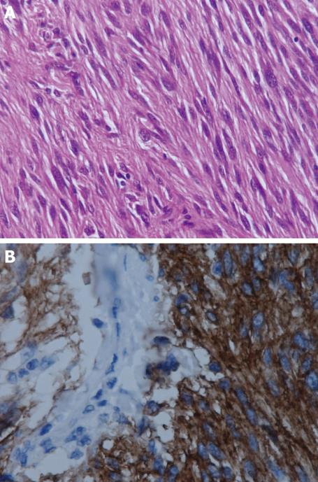
Figure 3 Histological findings and immunohistochemical examination.
A: Hematoxylin and eosin staining showed a spindle-cell morphology (original magnification 200 ×), B: An immunohistochemical examination revealed that the tumor cells were diffuse positive for KIT (original magnification 200 ×).
- Citation: Hiraki M, Kitajima Y, Ohtsuka T, Kai K, Miyake S, Koga Y, Mori D, Noshiro H, Tokunaga O, Miyazaki K. Immunohistochemical and molecular genetic analyses of multiple sporadic gastrointestinal stromal tumors. World J Gastrointest Oncol 2010; 2(9): 364-368
- URL: https://www.wjgnet.com/1948-5204/full/v2/i9/364.htm
- DOI: https://dx.doi.org/10.4251/wjgo.v2.i9.364


