Copyright
©2010 Baishideng.
World J Gastrointest Oncol. Jul 15, 2010; 2(7): 307-310
Published online Jul 15, 2010. doi: 10.4251/wjgo.v2.i7.307
Published online Jul 15, 2010. doi: 10.4251/wjgo.v2.i7.307
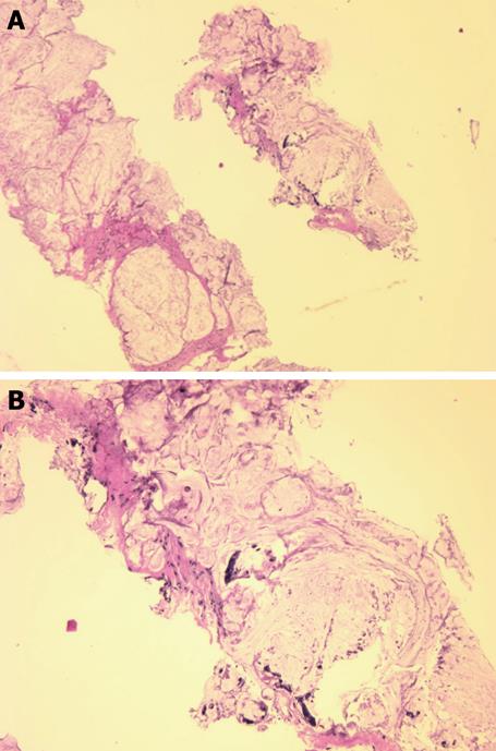
Figure 1 Needle biopsy fragments showing mucinous adenocarcinoma (HE stain).
A: Low power photomicrograph above shows fragments of infiltrating cancer; B: High power photomicrograph below shows individual cancer cells or clusters of cancer cells within mucus.
- Citation: Freeman HJ, Perry T, Webber DL, Chang SD, Loh MY. Mucinous carcinoma in Crohn’s disease originating in a fistulous tract. World J Gastrointest Oncol 2010; 2(7): 307-310
- URL: https://www.wjgnet.com/1948-5204/full/v2/i7/307.htm
- DOI: https://dx.doi.org/10.4251/wjgo.v2.i7.307


