
















Copyright: ©Author(s) 2026.
World J Gastrointest Oncol. Aug 15, 2026; 18(8): 121799
Published online Aug 15, 2026. doi: 10.4251/wjgo.v18.i8.121799
Published online Aug 15, 2026. doi: 10.4251/wjgo.v18.i8.121799
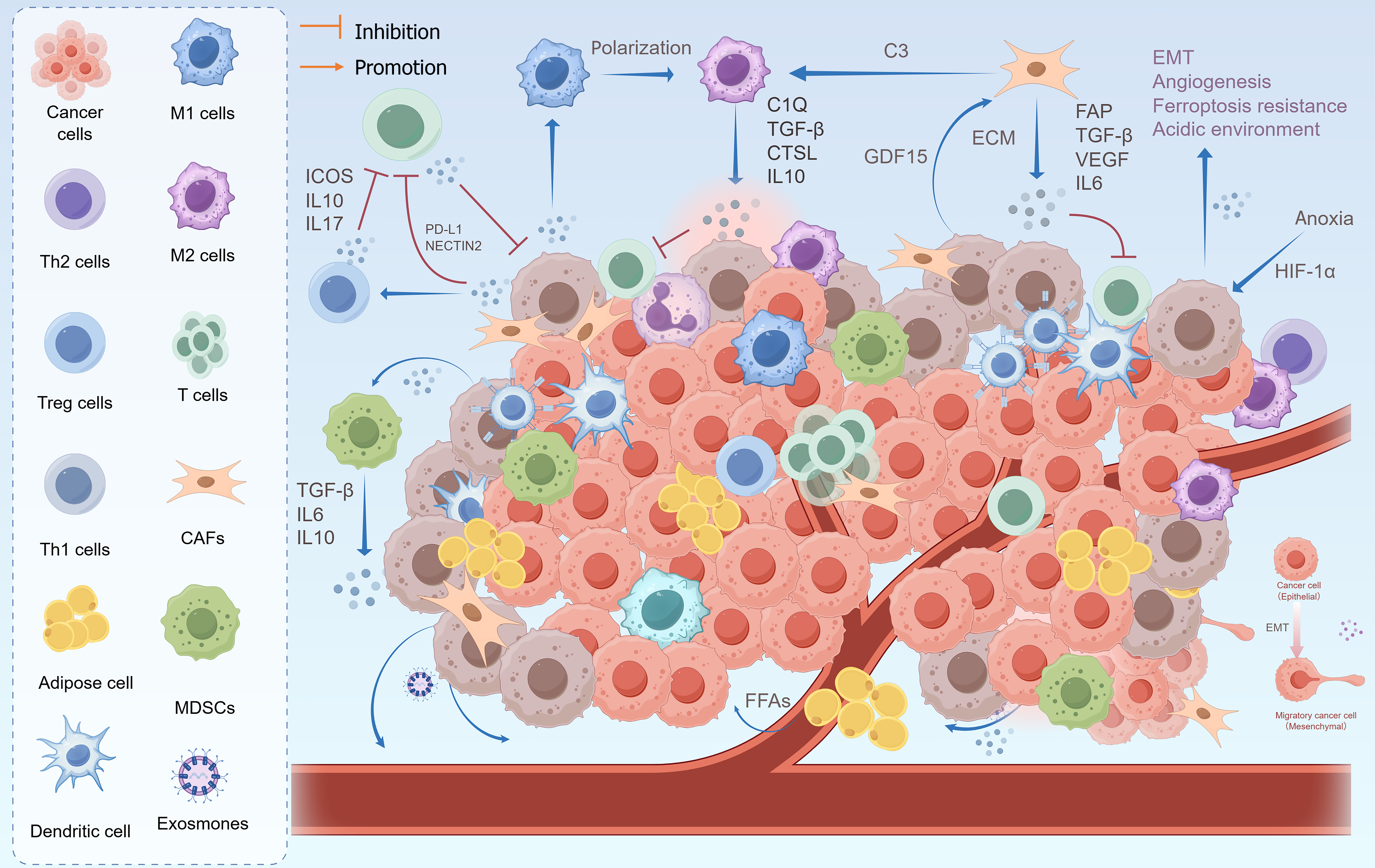
Figure 3 Outline of tumor microenvironment in peritoneal metastasis of gastric cancer.
M1 macrophages, dendritic cells, and T cells are innate immune cells that fight tumors. Gastric cancer cells induce effector T cells into an exhausted state by expressing relevant immune checkpoints, recruiting and promoting the polarization of M2 macrophages, and modifying regulatory T cell and myeloid-derived suppressor cells to suppress immune responses. Immune cells normally secrete cytokines to suppress tumor growth, however, suppressor T cells cannot produce these cytokines. In addition, gastric cancer cells induce the differentiation of cancer-associated fibroblasts and promote the maturation of preadipocytes, shaping the aggressive phenotype and immune tolerance of tumor cells. Anoxia is a core feature of peritoneal metastasis, which can drive epithelial-mesenchymal transition, angiogenesis, ferroptosis resistance, and acidic tumor microenvironment. EMT: Epithelial-mesenchymal transition; ICOS: Inducible T-cell co-stimulator; IL: Interleukin; PD-L1: Programmed death ligand 1; TGF: Transforming growth factor; CTSL: Cathepsin L; GDF15: Growth differentiation factor 15; ECM: Extracellular matrix; FAP: Fibronectin attachment protein; VEGF: Vascular endothelial growth factor; HIF-1α: Hypoxia-inducible factor 1alpha; CAFs: Cancer-associated fibroblasts; MDSC: Myeloid-derived suppressor cell; FFAs: Free fatty acids.
- Citation: Yan HQ, Lai QQ, Huang MW, Wang TA, Wu LC, Qin YZ. Advances in molecular mechanism of gastric cancer peritoneal metastasis. World J Gastrointest Oncol 2026; 18(8): 121799
- URL: https://www.wjgnet.com/1948-5204/full/v18/i8/121799.htm
- DOI: https://dx.doi.org/10.4251/wjgo.v18.i8.121799