
















Copyright: ©Author(s) 2026.
World J Gastrointest Oncol. Aug 15, 2026; 18(8): 121799
Published online Aug 15, 2026. doi: 10.4251/wjgo.v18.i8.121799
Published online Aug 15, 2026. doi: 10.4251/wjgo.v18.i8.121799
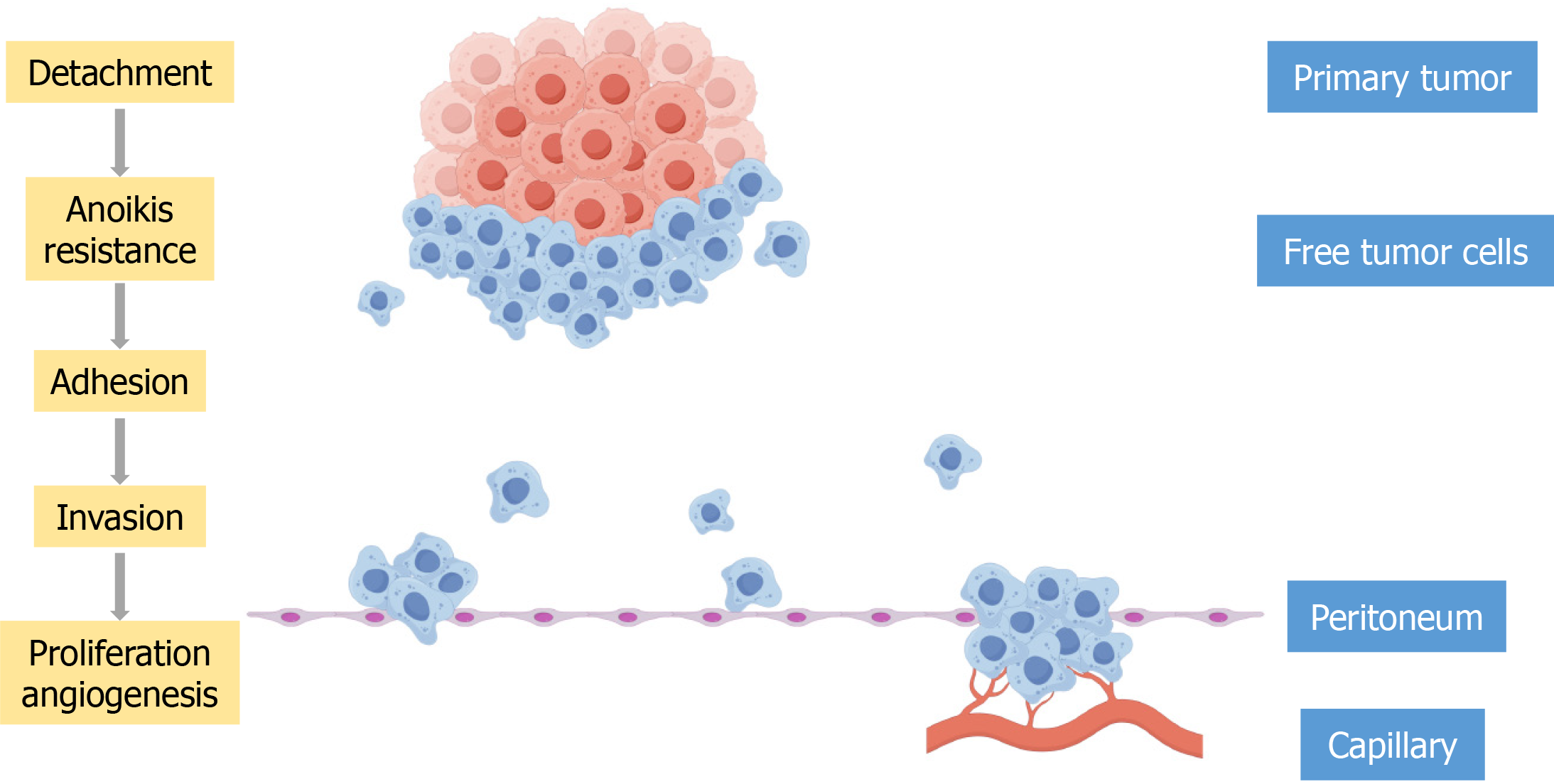
Figure 1 The stepwise process of gastric cancer peritoneal metastasis.
This schematic illustrates the sequential steps by which gastric cancer cells disseminate from the primary tumor and establish metastatic lesions in the peritoneum. The process involves six critical phases: (1) Shedding: Gastric cancer cells detach from the primary tumor by downregulating intercellular adhesion molecules; (2) Anoikis resistance: Detached cells survive in the peritoneal fluid by activating pro-survival pathways; (3) Adhesion: Free-floating cells adhere to the peritoneal mesothelial layer via adhesion molecules; (4) Invasion: Adhered cells secrete matrix metalloproteinases and cathepsins to degrade the extracellular matrix, invading the subperitoneal space; (5) Proliferation: Invaded cells proliferate, forming micrometastases; and (6) Angiogenesis: The growing metastatic foci secrete angiogenic factors (e.g., vascular endothelial growth factor), inducing new blood vessel formation to supply nutrients and oxygen.
- Citation: Yan HQ, Lai QQ, Huang MW, Wang TA, Wu LC, Qin YZ. Advances in molecular mechanism of gastric cancer peritoneal metastasis. World J Gastrointest Oncol 2026; 18(8): 121799
- URL: https://www.wjgnet.com/1948-5204/full/v18/i8/121799.htm
- DOI: https://dx.doi.org/10.4251/wjgo.v18.i8.121799