
















Copyright: ©Author(s) 2026.
World J Gastrointest Oncol. Aug 15, 2026; 18(8): 121073
Published online Aug 15, 2026. doi: 10.4251/wjgo.121073
Published online Aug 15, 2026. doi: 10.4251/wjgo.121073
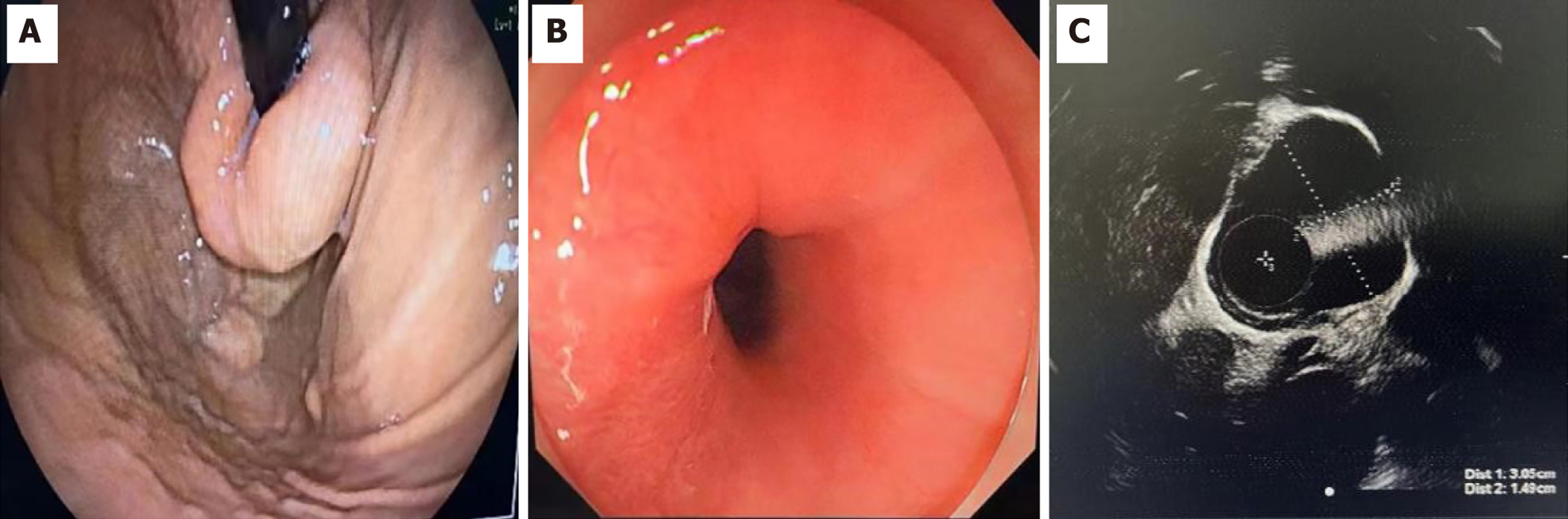
Figure 1 Endoscopic and endoscopic ultrasonographic views of the lesion.
A: Gastroscopic view of the lesion from the gastric fundus side; B: Gastroscopic view of the lesion from the esophageal side; C: Endoscopic ultrasonographic view of the lesion showing a hypoechoic mass originating from the muscularis propria layer.
- Citation: Mai WJ, Miao W, Ma YM, Zhou HH, Yuan S. Submucosal tunnel docking endoscopic resection for a large gastroesophageal junction leiomyoma: A case report. World J Gastrointest Oncol 2026; 18(8): 121073
- URL: https://www.wjgnet.com/1948-5204/full/v18/i8/121073.htm
- DOI: https://dx.doi.org/10.4251/wjgo.121073