
















Copyright: ©Author(s) 2026.
World J Gastrointest Oncol. Aug 15, 2026; 18(8): 120982
Published online Aug 15, 2026. doi: 10.4251/wjgo.v18.i8.120982
Published online Aug 15, 2026. doi: 10.4251/wjgo.v18.i8.120982
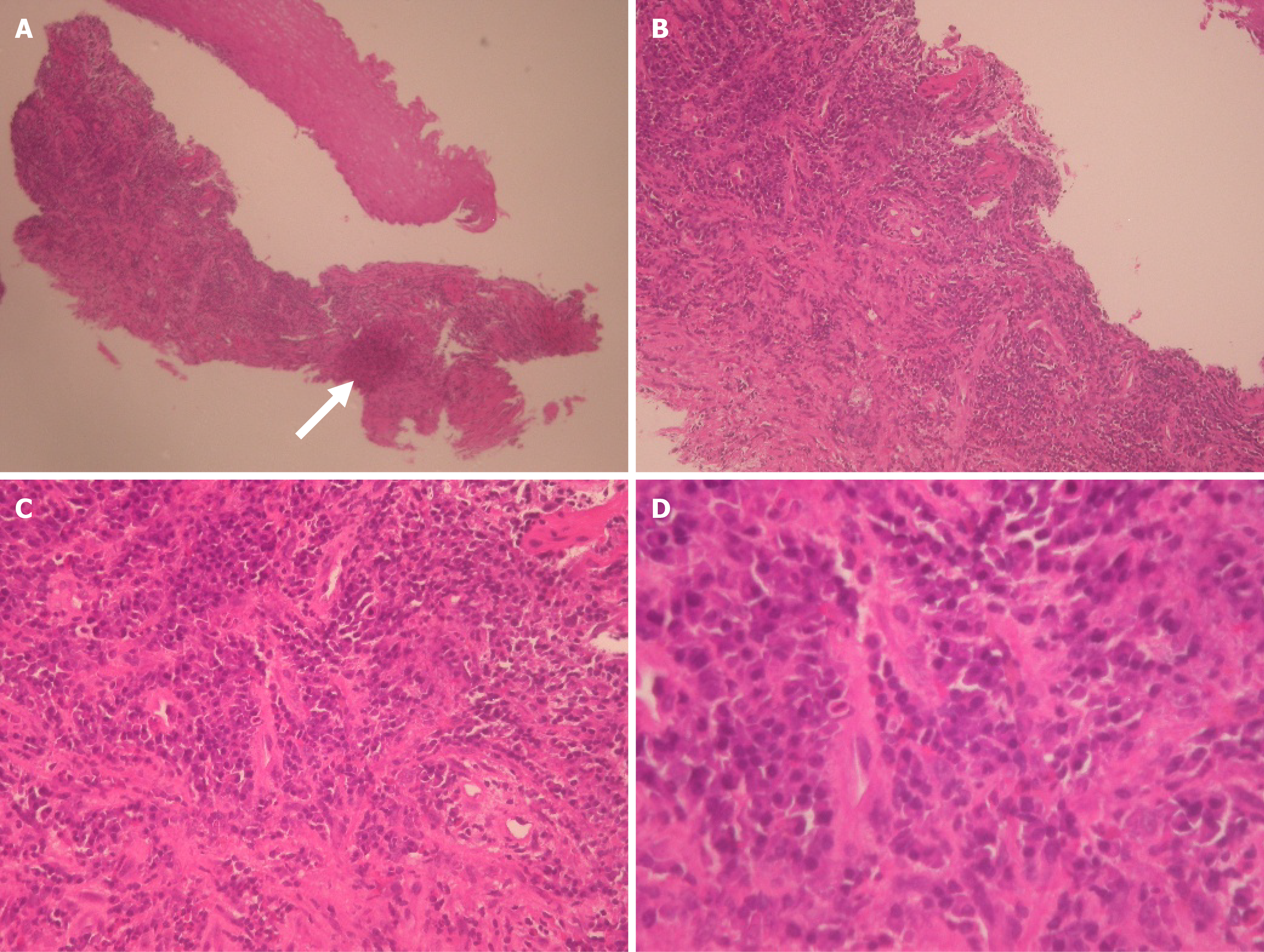
Figure 6 Histopathological findings after treatment.
Hematoxylin and eosin-stained sections from post-treatment endoscopic biopsy specimens were evaluated. A: Low-power view (× 40): Post-treatment biopsy showing preserved architecture of the esophageal mucosa with evidence of chronic inflammatory cell infiltration in the stroma. The arrow indicates a representative area for detailed evaluation; B: Medium-power view (× 100): Magnified view of the arrow-indicated area in (A). The squamous epithelium demonstrates regenerative changes with restoration of normal stratification and maturation. No evidence of expansile epithelial proliferation or tumor nests is observed; C: High-power view (× 200): Further magnification of the same region. The epithelial cells lack cytologic atypia, with normal nuclear-to-cytoplasmic ratios. No dysplastic or malignant features are identified; D: High-power view (× 400): Highest magnification of the arrow-indicated area. Post-treatment endoscopic biopsy specimen demonstrates subepithelial connective tissue with prominent inflammatory infiltrates composed predominantly of neutrophils and lymphocytes. No cytologic atypia or malignant squamous cells are identified in the examined field. These findings support the presence of a pathological complete response at the primary tumor site.
- Citation: Cheng CW, Li WT, Lee YT, Kam KH, Tsang YW. Long-term complete response in stage IVa esophageal squamous cell carcinoma treated with chemoradiotherapy and modulated electro-hyperthermia: A case report. World J Gastrointest Oncol 2026; 18(8): 120982
- URL: https://www.wjgnet.com/1948-5204/full/v18/i8/120982.htm
- DOI: https://dx.doi.org/10.4251/wjgo.v18.i8.120982