
















Copyright: ©Author(s) 2026.
World J Gastrointest Oncol. Aug 15, 2026; 18(8): 120982
Published online Aug 15, 2026. doi: 10.4251/wjgo.v18.i8.120982
Published online Aug 15, 2026. doi: 10.4251/wjgo.v18.i8.120982
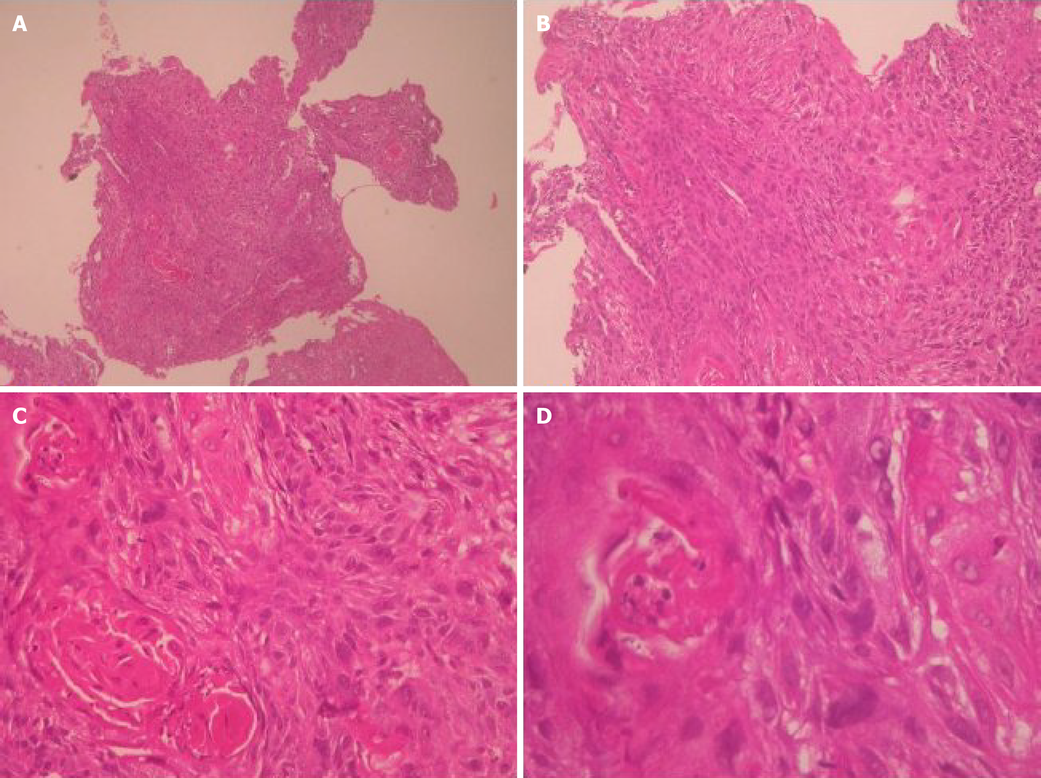
Figure 3 Histopathological features of esophageal squamous cell carcinoma prior to treatment.
Hematoxylin and eosin-stained sections from endoscopic biopsy specimens were examined. A: Low-power view (× 40): Multiple small, fragmented pieces of esophageal mucosa are shown. The limited tissue size (0.3 cm × 0.2 cm × 0.1 cm) constrains evaluation of deeper structures; B: Medium-power view (× 100): The squamous epithelium exhibits complete loss of normal stratification and maturation, with atypical cells occupying the entire layer; C: High-power view (× 200): Neoplastic squamous cells exhibit prominent hyperchromatic nuclei and pleomorphism, involving the full thickness of the epithelium. These features are diagnostic of at least high-grade squamous intraepithelial neoplasia (squamous cell carcinoma in situ); D: High-power view (× 400): Focal areas show keratin pearl formation, supporting squamous differentiation. While subepithelial stromal invasion is suspected, a definitive diagnosis of invasive carcinoma is precluded by the limited depth of the specimen. These findings are consistent with squamous cell carcinoma in situ with features suspicious for early invasion.
- Citation: Cheng CW, Li WT, Lee YT, Kam KH, Tsang YW. Long-term complete response in stage IVa esophageal squamous cell carcinoma treated with chemoradiotherapy and modulated electro-hyperthermia: A case report. World J Gastrointest Oncol 2026; 18(8): 120982
- URL: https://www.wjgnet.com/1948-5204/full/v18/i8/120982.htm
- DOI: https://dx.doi.org/10.4251/wjgo.v18.i8.120982