
















Copyright: ©Author(s) 2026.
World J Gastrointest Oncol. Aug 15, 2026; 18(8): 120982
Published online Aug 15, 2026. doi: 10.4251/wjgo.v18.i8.120982
Published online Aug 15, 2026. doi: 10.4251/wjgo.v18.i8.120982
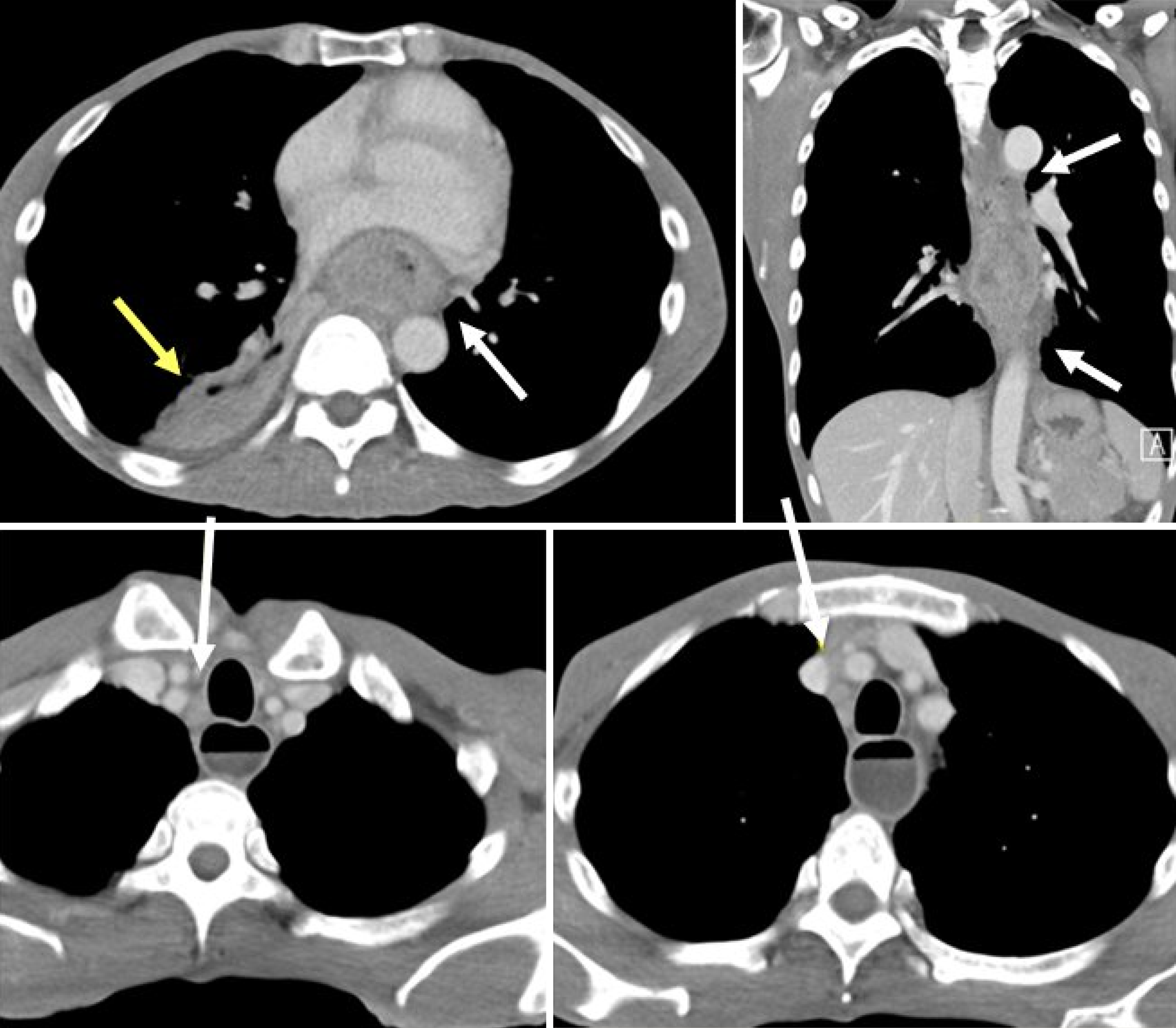
Figure 1 Initial chest computed tomography showing esophageal tumor and pneumonia.
Contrast-enhanced axial computed tomography scan demonstrates a large soft-tissue mass involving the mid- to lower-third esophagus (white arrow), with associated paratracheal and paraesophageal lymphadenopathy. Consolidation and abscess formation suggestive of pneumonia (yellow arrow) with a suspicious tracheoesophageal fistula are also observed.
- Citation: Cheng CW, Li WT, Lee YT, Kam KH, Tsang YW. Long-term complete response in stage IVa esophageal squamous cell carcinoma treated with chemoradiotherapy and modulated electro-hyperthermia: A case report. World J Gastrointest Oncol 2026; 18(8): 120982
- URL: https://www.wjgnet.com/1948-5204/full/v18/i8/120982.htm
- DOI: https://dx.doi.org/10.4251/wjgo.v18.i8.120982