
















Copyright: ©Author(s) 2026.
World J Gastrointest Oncol. Aug 15, 2026; 18(8): 119679
Published online Aug 15, 2026. doi: 10.4251/wjgo.119679
Published online Aug 15, 2026. doi: 10.4251/wjgo.119679
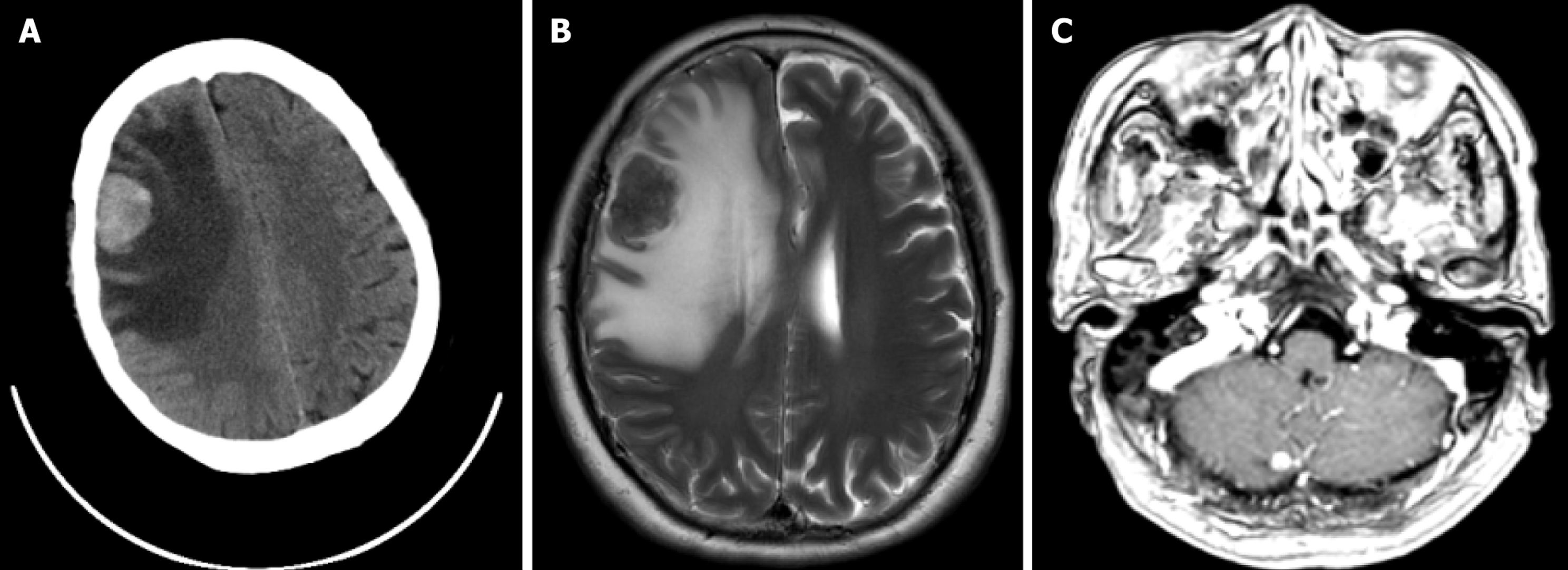
Figure 1 Head computed tomography and magnetic resonance imaging examinations of the patient.
A: Head computed tomography revealed an abnormal density focus in the right frontal lobe; B: Brain magnetic resonance imaging demonstrated a space-occupying lesion in the right frontal cortex, measuring approximately 3.1 cm × 2.4 cm; C: A follow-up brain magnetic resonance imaging scan prior to radiotherapy showed a newly developed small nodular lesion, approximately 0.6 cm in diameter, in the right cerebellar hemisphere.
- Citation: Xu JL, Ge ZW, Wu CW, Wang YH, Mao JD. Multidisciplinary care for colorectal cancer brain metastases with headache as initial symptom: A case report and literature review. World J Gastrointest Oncol 2026; 18(8): 119679
- URL: https://www.wjgnet.com/1948-5204/full/v18/i8/119679.htm
- DOI: https://dx.doi.org/10.4251/wjgo.119679