
















Copyright: ©Author(s) 2026.
World J Gastrointest Oncol. Jul 15, 2026; 18(7): 120564
Published online Jul 15, 2026. doi: 10.4251/wjgo.120564
Published online Jul 15, 2026. doi: 10.4251/wjgo.120564
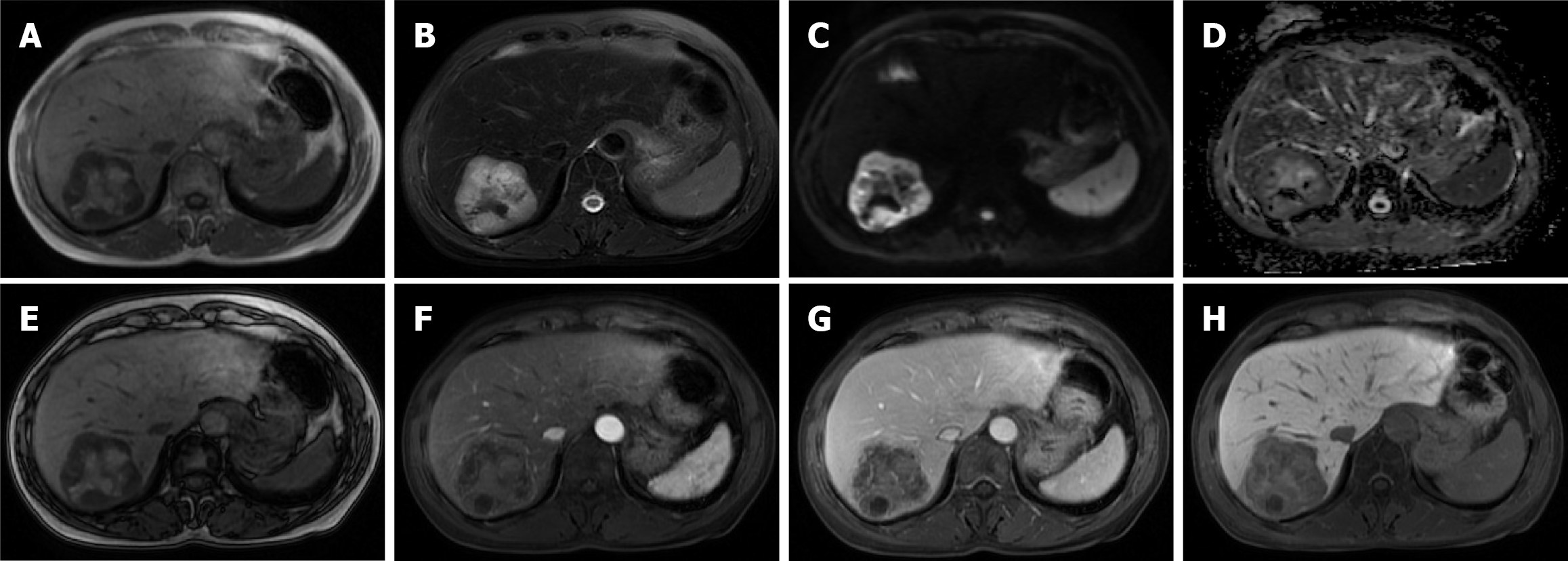
Figure 2 Magnetic resonance imaging revealed a heterogeneous signal mass in segment VII of the liver, measuring approximately 64 mm × 62 mm.
A: T1-weighted image shows predominant hypointensity with internal iso- to hyperintense foci; B: T2-weighted image shows predominant hyperintensity with internal hypointense areas; C and D: Diffusion-weighted imaging (C) and apparent diffusion coefficient (D) demonstrate peripheral restricted diffusion, indicating high cellularity at the viable tumor rim; E: Opposed-phase imaging shows no significant signal loss, arguing against microscopic fat; F and G: Contrast-enhanced imaging reveals patchy, heterogeneous enhancement; H: Hepatobiliary phase shows distinct hypointensity relative to background liver, indicating a lack of functioning hepatocytes.
- Citation: Xing HQ, Mo TM, He J. Primary hepatic adenoid cystic carcinoma: A case report and review of literature. World J Gastrointest Oncol 2026; 18(7): 120564
- URL: https://www.wjgnet.com/1948-5204/full/v18/i7/120564.htm
- DOI: https://dx.doi.org/10.4251/wjgo.120564