
















Copyright: ©Author(s) 2026.
World J Gastrointest Oncol. Jul 15, 2026; 18(7): 120564
Published online Jul 15, 2026. doi: 10.4251/wjgo.120564
Published online Jul 15, 2026. doi: 10.4251/wjgo.120564
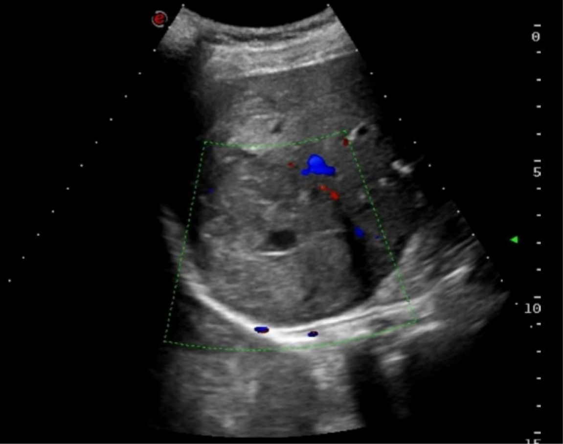
Figure 1 Ultrasound revealed a cystic-solid mass measuring approximately 68 mm × 59 mm in the right lobe of the liver.
A central fluid-dark area with poor internal sound transmission (indicative of necrosis) and peripheral blood flow signals were observed. These features are non-specific and may mimic benign lesions such as a liver abscess.
- Citation: Xing HQ, Mo TM, He J. Primary hepatic adenoid cystic carcinoma: A case report and review of literature. World J Gastrointest Oncol 2026; 18(7): 120564
- URL: https://www.wjgnet.com/1948-5204/full/v18/i7/120564.htm
- DOI: https://dx.doi.org/10.4251/wjgo.120564