
















Copyright: ©Author(s) 2026.
World J Gastrointest Oncol. Jul 15, 2026; 18(7): 118212
Published online Jul 15, 2026. doi: 10.4251/wjgo.118212
Published online Jul 15, 2026. doi: 10.4251/wjgo.118212
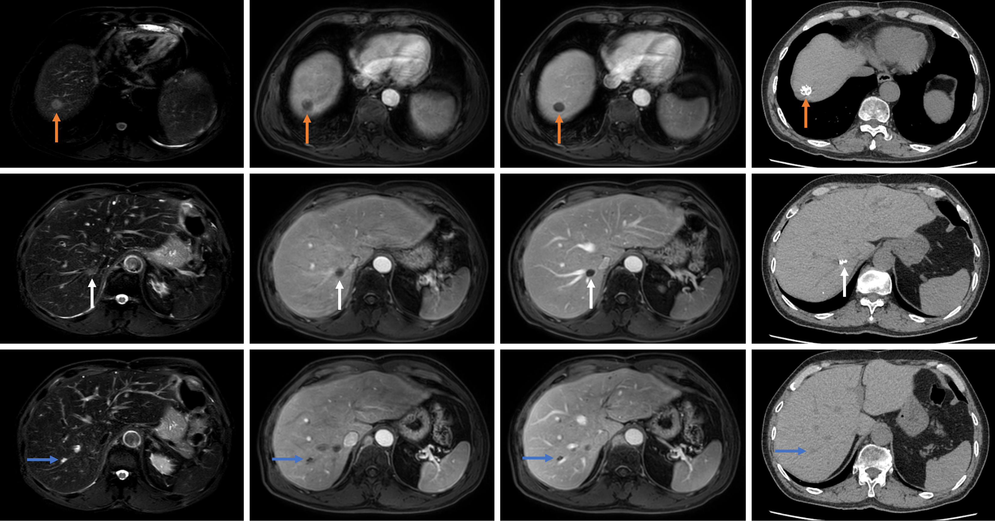
Figure 4 At the 42-month postoperative follow-up, the three occult micrometastases showed no significant blood flow or enhancement on magnetic resonance imaging during both arterial and portal venous phases, with gradual shrinkage.
Computed tomography confirmed satisfactory lipiodol deposition within some tumors. Orange arrow: Dorsal portion of hepatic segment 8; White arrow: Paracaval portion lesion; Blue arrow: Segment 7 lesion.
- Citation: Yang CW, Zhang RX, Zheng XL, Zhu L, Zheng Z, Wu XH. Long-term survival after combined hepatectomy and colectomy for ruptured combined hepatocellular-cholangiocarcinoma with direct colonic invasion: A case report. World J Gastrointest Oncol 2026; 18(7): 118212
- URL: https://www.wjgnet.com/1948-5204/full/v18/i7/118212.htm
- DOI: https://dx.doi.org/10.4251/wjgo.118212