
















Copyright: ©Author(s) 2026.
World J Gastrointest Oncol. Jul 15, 2026; 18(7): 118212
Published online Jul 15, 2026. doi: 10.4251/wjgo.118212
Published online Jul 15, 2026. doi: 10.4251/wjgo.118212
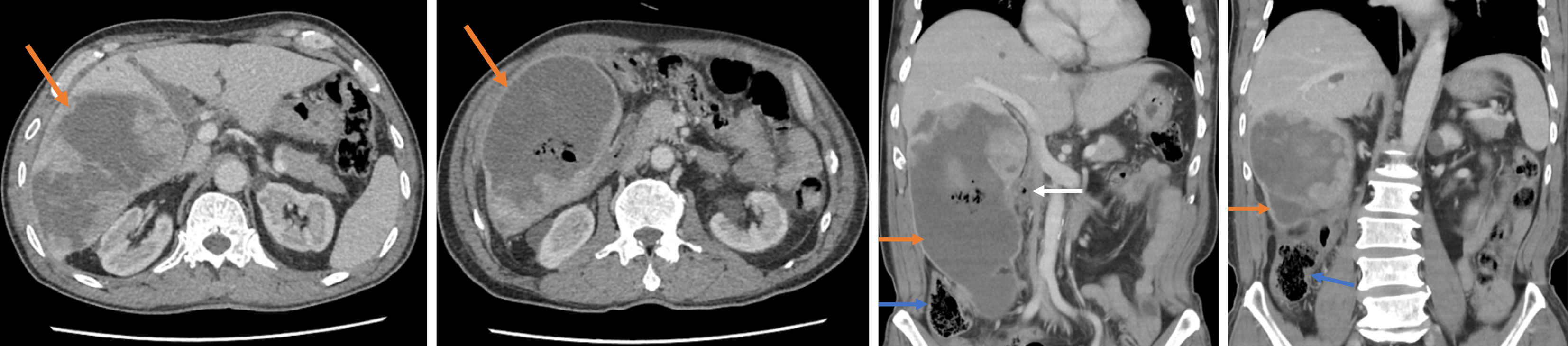
Figure 1 Abdominal contrast-enhanced computed tomography showed a large heterogeneous right hepatic lobe mass with internal high-density material, gas shadows, heterogeneous enhancement, and blurred adjacent fat planes.
Orange arrow: Liver mass; White arrow: Duodenum; Blue arrow: Colon.
- Citation: Yang CW, Zhang RX, Zheng XL, Zhu L, Zheng Z, Wu XH. Long-term survival after combined hepatectomy and colectomy for ruptured combined hepatocellular-cholangiocarcinoma with direct colonic invasion: A case report. World J Gastrointest Oncol 2026; 18(7): 118212
- URL: https://www.wjgnet.com/1948-5204/full/v18/i7/118212.htm
- DOI: https://dx.doi.org/10.4251/wjgo.118212