
















Copyright: ©Author(s) 2026.
World J Gastrointest Oncol. Jun 15, 2026; 18(6): 118708
Published online Jun 15, 2026. doi: 10.4251/wjgo.v18.i6.118708
Published online Jun 15, 2026. doi: 10.4251/wjgo.v18.i6.118708
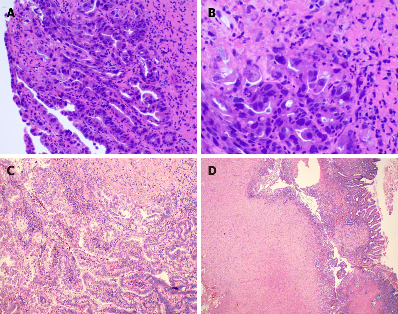
Figure 3 Hematoxylin and eosin staining was performed on the tumor tissue.
A and B: The structure of the gastric mucosal glands was disordered, presenting a sieve-like fusion-like change. The glandular epithelial cells are significantly abnormal, manifested as enlarged nuclei, increased nucleoplasmic ratio, disordered cell arrangement, and loss of polarity; C and D: The cancer cells are arranged in an irregular glandular and nest-like pattern, with obvious cell atypia and distinct mitotic signs. The tumor infiltrated into the submucosa and invaded the surrounding leiomyoma tissue. The measured depth of infiltration was approximately 4.26 mm.
- Citation: Wang QJ, Gao JN, Wu PT, Lu GR, Xu XZ. Moderately to poorly differentiated adenocarcinoma invading a leiomyoma: A case report. World J Gastrointest Oncol 2026; 18(6): 118708
- URL: https://www.wjgnet.com/1948-5204/full/v18/i6/118708.htm
- DOI: https://dx.doi.org/10.4251/wjgo.v18.i6.118708