
















Copyright: ©Author(s) 2026.
World J Gastrointest Oncol. Jun 15, 2026; 18(6): 118708
Published online Jun 15, 2026. doi: 10.4251/wjgo.v18.i6.118708
Published online Jun 15, 2026. doi: 10.4251/wjgo.v18.i6.118708
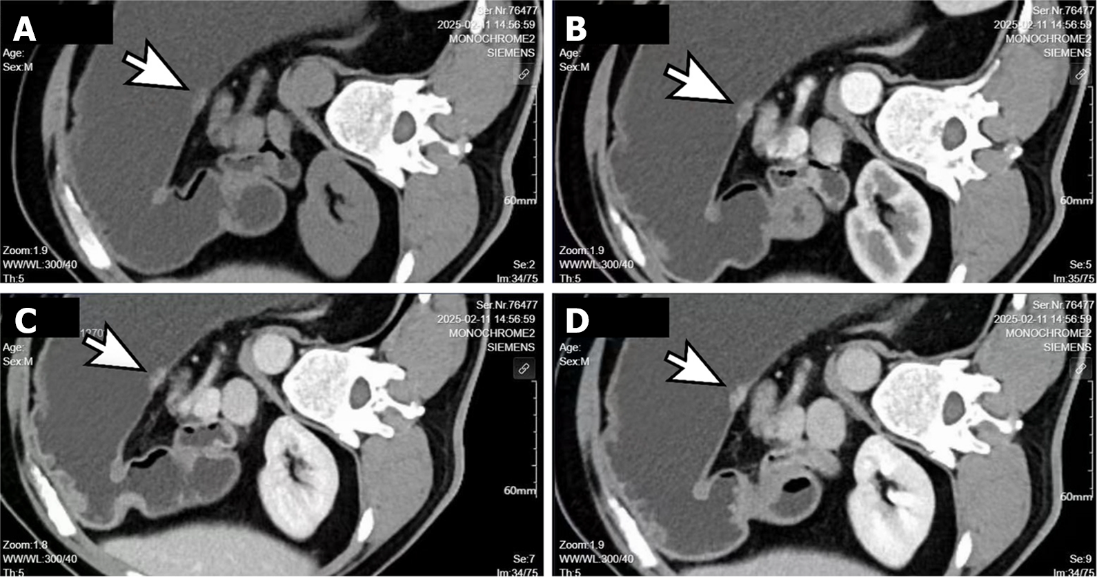
Figure 2 Enhanced gastric computed tomography.
The posterior wall of the lower gastric body shows focal nodular thickening measuring approximately 16 mm × 8 mm. On contrast-enhanced imaging, the lesion demonstrates progressive, marked enhancement with heterogeneous filling. The lesion protrudes into the gastric lumen with localized surface indentation. The serosal margin of the lesion appears smooth. No significant perigastric lymphadenopathy is noted. A: Non-contrast; B: Arterial phase; C: Venous phase; D: Delayed phase. Arrow: Location of the lesion.
- Citation: Wang QJ, Gao JN, Wu PT, Lu GR, Xu XZ. Moderately to poorly differentiated adenocarcinoma invading a leiomyoma: A case report. World J Gastrointest Oncol 2026; 18(6): 118708
- URL: https://www.wjgnet.com/1948-5204/full/v18/i6/118708.htm
- DOI: https://dx.doi.org/10.4251/wjgo.v18.i6.118708