
















Copyright: ©Author(s) 2026.
World J Gastrointest Oncol. Jun 15, 2026; 18(6): 118708
Published online Jun 15, 2026. doi: 10.4251/wjgo.v18.i6.118708
Published online Jun 15, 2026. doi: 10.4251/wjgo.v18.i6.118708
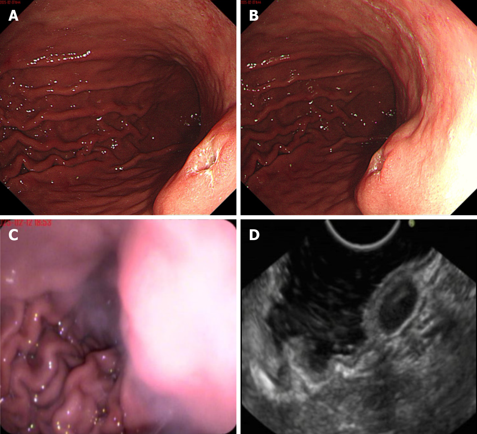
Figure 1 Endoscopic findings.
A and B: Visible under white light: A hemispherical mucosal elevation is observed on the posterior wall of the gastric body, measuring approximately 1.5 cm × 2.0 cm in diameter. The surface is depressed with ulceration, covered by a white coating, and the surrounding mucosa appears congested and edematous; C and D: Endoscopic ultrasound findings: The gastric body lesion was hypoechoic, ellipsoid-like, with a still homogeneous internal echo and partially visible hypoechoic, and the size of the slice was about 1.0 cm × 1.0 cm, with a still clear border, originating in the intrinsic muscularis propria layer.
- Citation: Wang QJ, Gao JN, Wu PT, Lu GR, Xu XZ. Moderately to poorly differentiated adenocarcinoma invading a leiomyoma: A case report. World J Gastrointest Oncol 2026; 18(6): 118708
- URL: https://www.wjgnet.com/1948-5204/full/v18/i6/118708.htm
- DOI: https://dx.doi.org/10.4251/wjgo.v18.i6.118708