
















Copyright: ©Author(s) 2026.
World J Gastrointest Oncol. Jun 15, 2026; 18(6): 117697
Published online Jun 15, 2026. doi: 10.4251/wjgo.v18.i6.117697
Published online Jun 15, 2026. doi: 10.4251/wjgo.v18.i6.117697
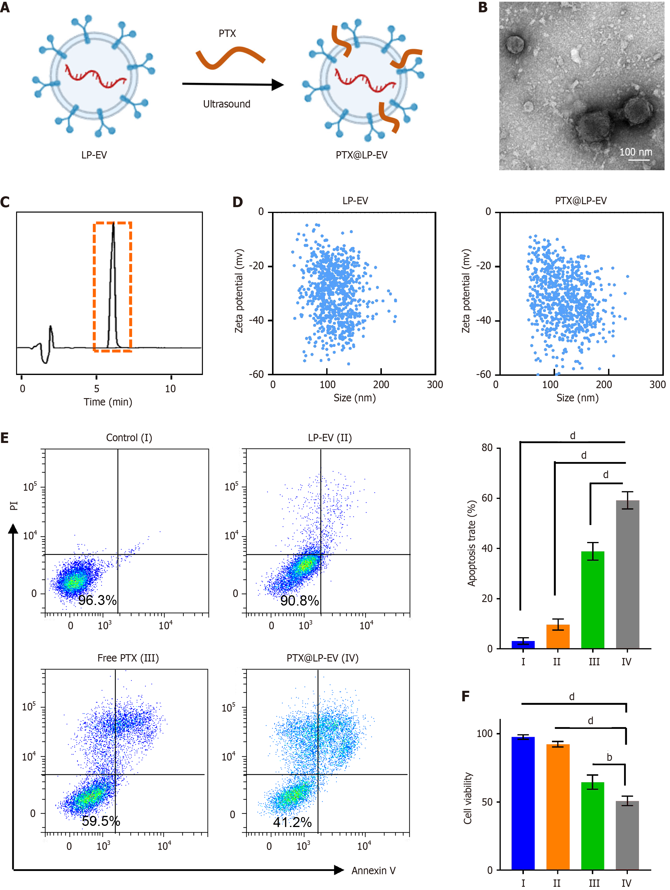
Figure 3 Preparation and in vitro anti-tumor evaluation of paclitaxel-loaded low-pH preconditioned macrophage-derived extracellular vesicles.
A: Schematic illustrating the preparation of paclitaxel (PTX)-loaded low-pH preconditioned macrophage-derived extracellular vesicles (LP-EV) (PTX@LP-EV) via sonication; B: Representative transmission electron microscopy image of PTX@LP-EV. Scale bar: 100 nm; C: High-performance liquid chromatography chromatograms of PTX@LP-EV lysate; D: Nanoparticle tracking analysis showing the size distribution and zeta potential of LP-EV and PTX@LP-EV; E: Flow cytometry analysis of apoptosis in MKN45 cells after treatment with PBS, LP-EV, free PTX, or PTX@LP-EV. Quantitative data are shown on the right; F: Viability of MKN45 cells after treatment with the indicated formulations, as assessed by Cell Counting Kit-8 assay. Data in (E and F) represent means ± SD. Statistical significance was tested using one-way ANOVA with multiple comparison tests, bP < 0.01, dP < 0.0001. LP-EV: Low-pH preconditioned macrophage-derived extracellular vesicle; PI: Propidium iodide; PTX: Paclitaxel.
- Citation: Wang JH, Hu ML, Shi M, Ma JL, Yang J, Wang YG. Low-pH preconditioned macrophage-derived extracellular vesicles enable targeted and enhanced paclitaxel therapy in gastric cancer. World J Gastrointest Oncol 2026; 18(6): 117697
- URL: https://www.wjgnet.com/1948-5204/full/v18/i6/117697.htm
- DOI: https://dx.doi.org/10.4251/wjgo.v18.i6.117697