
















Copyright: ©Author(s) 2026.
World J Gastrointest Oncol. Jun 15, 2026; 18(6): 117697
Published online Jun 15, 2026. doi: 10.4251/wjgo.v18.i6.117697
Published online Jun 15, 2026. doi: 10.4251/wjgo.v18.i6.117697
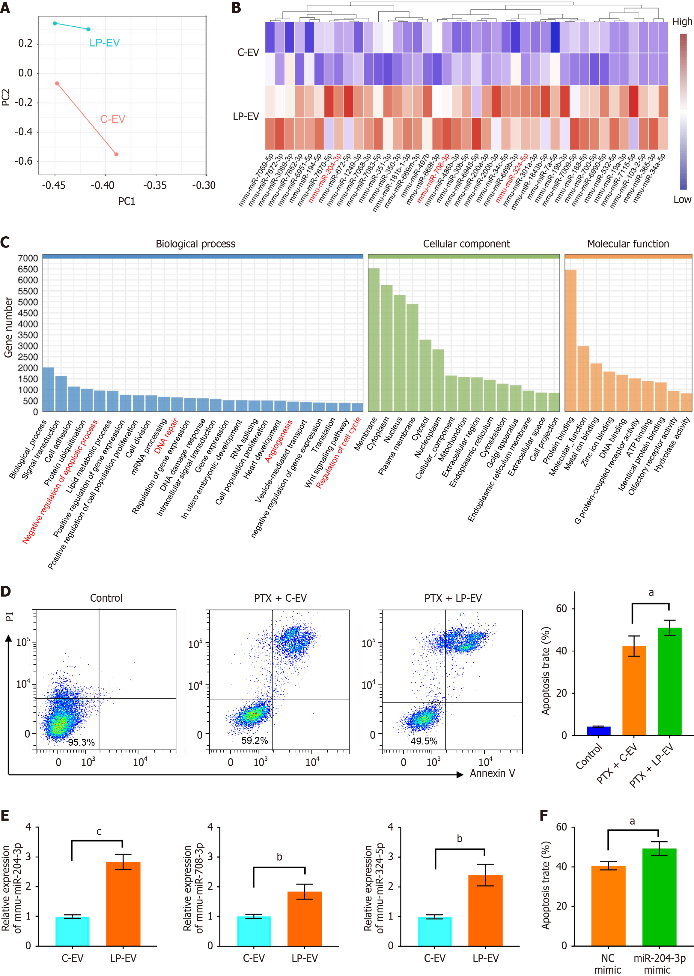
Figure 2 miRNA profiling and functional analysis of low-pH preconditioned macrophage-derived extracellular vesicles.
A: Principal component analysis of miRNA expression profiles from control-extracellular vesicles (C-EV) and low-pH preconditioned macrophage-derived EVs (LP-EV); B: Heatmap displaying upregulated miRNAs in LP-EV compared to C-EV (|log2 fold change| > 1, P < 0.05); C: Gene Ontology enrichment analysis of predicted target genes for miRNAs upregulated in LP-EV; D: Flow cytometry analysis of apoptosis in MKN45 cells treated with PBS, paclitaxel (PTX) combined with C-EV, or PTX combined with LP-EV. The right panel shows the quantitative analysis of apoptotic cell percentages; E: Quantitative real-time PCR validation of selected miRNAs (miR-204-3p, miR-708-3p, and miR-324-5p) in LP-EV compared with C-EV; F: Quantitative analysis of apoptotic cell populations in MKN45 cells transfected with negative control mimic or miR-204-3p mimic, followed by treatment with PTX in combination with C-EV. Data in (D-F) represent means ± SD. Statistical significance was tested using unpaired t test (E and F) or one-way ANOVA with multiple comparison tests (D), aP < 0.05, bP < 0.01, cP < 0.001. LP-EV: Low-pH preconditioned macrophage-derived extracellular vesicle; C-EV: Control extracellular vesicle; PTX: Paclitaxel; PI: Propidium iodide; NC: Negative control.
- Citation: Wang JH, Hu ML, Shi M, Ma JL, Yang J, Wang YG. Low-pH preconditioned macrophage-derived extracellular vesicles enable targeted and enhanced paclitaxel therapy in gastric cancer. World J Gastrointest Oncol 2026; 18(6): 117697
- URL: https://www.wjgnet.com/1948-5204/full/v18/i6/117697.htm
- DOI: https://dx.doi.org/10.4251/wjgo.v18.i6.117697