
















Copyright: ©Author(s) 2026.
World J Gastrointest Oncol. Jun 15, 2026; 18(6): 117101
Published online Jun 15, 2026. doi: 10.4251/wjgo.v18.i6.117101
Published online Jun 15, 2026. doi: 10.4251/wjgo.v18.i6.117101
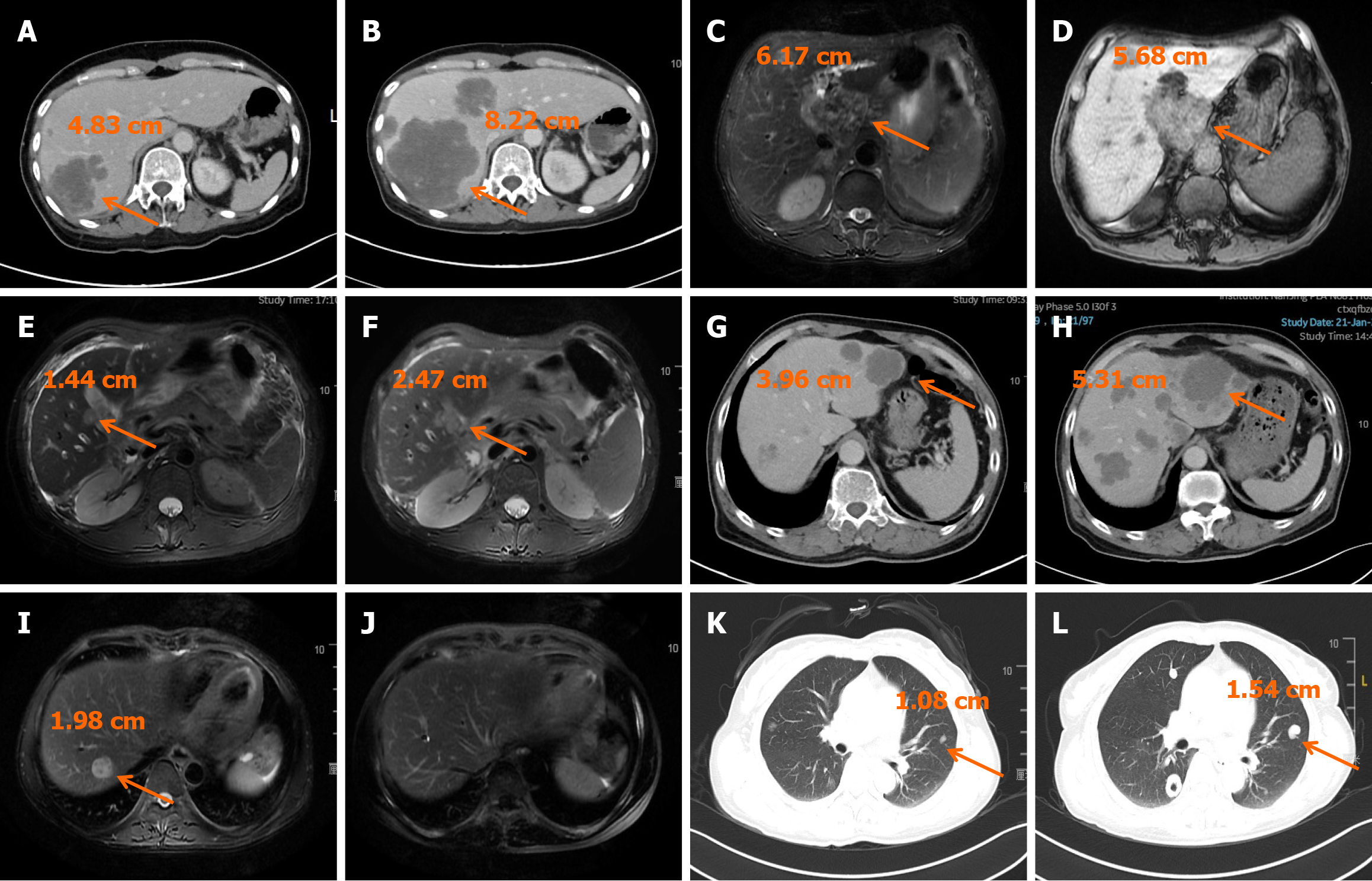
Figure 3 Imaging.
A: Patient 1, baseline; B: Patient 1, after 3 cycles; C: Patient 2, baseline; D: Patient 2, after 4 cycles; E: Patient 3, baseline; F: Patient 3, after 3 cycles; G: Patient 4, baseline; H: Patient 4, after 6 cycles; I: Patient 5, baseline; J: Patient 5, after 12 cycles; K: Patient 6, baseline; L: Patient 6, after 2 cycles. Orange arrows indicate the main lesions. Baseline refers to imaging at initiation of fruquintinib combined with sintilimab.
- Citation: Pan J, Li P, Zhao Y, Zhou YH, Zhao Y, Zhang TL, Chen YT, Chu XY. Efficacy and safety of fruquintinib plus sintilimab in third-line treatment of metastatic colorectal cancer: A single-center experience. World J Gastrointest Oncol 2026; 18(6): 117101
- URL: https://www.wjgnet.com/1948-5204/full/v18/i6/117101.htm
- DOI: https://dx.doi.org/10.4251/wjgo.v18.i6.117101