
















Copyright: ©Author(s) 2026.
World J Gastrointest Oncol. May 15, 2026; 18(5): 116776
Published online May 15, 2026. doi: 10.4251/wjgo.v18.i5.116776
Published online May 15, 2026. doi: 10.4251/wjgo.v18.i5.116776
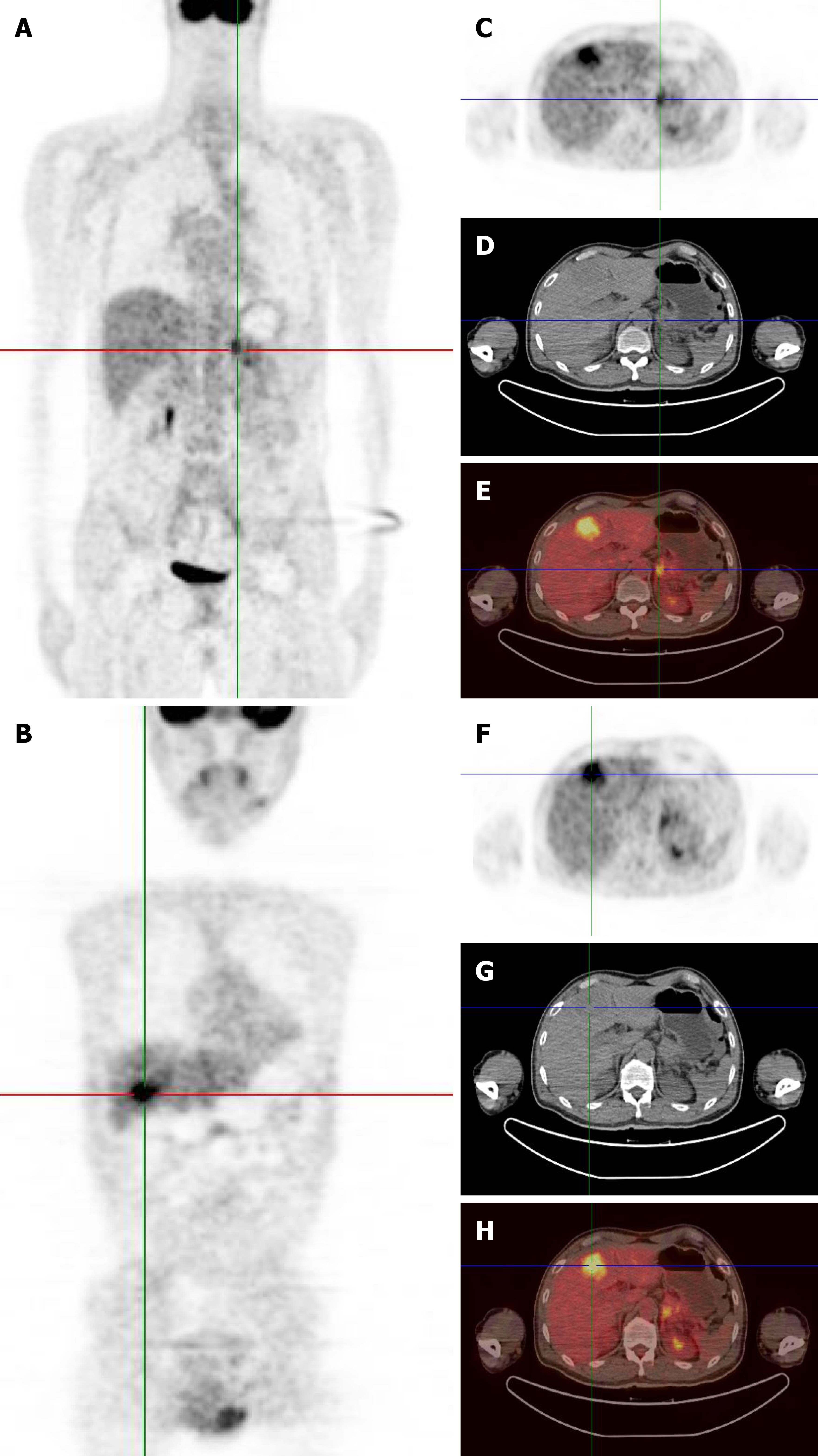
Figure 1 Preoperative 18F-fluorodeoxyglucose positron emission tomography/computed tomography imaging characteristics of the patient.
A: Preoperative 18F-fluorodeoxyglucose (18F-FDG) positron emission tomography/computed tomography (CT) maximum intensity projection images revealed intense uptake of 18F-FDG in the pancreatic body and tail (standardized uptake value maximum of 4.7); B: Preoperative 18F-FDG positron emission tomography/CT maximum intensity projection images revealed intense uptake of 18F-FDG in liver segment 4 (S4) (standardized uptake value maximum of 7.3); C: Positron emission tomography (PET) image showed a contrast-enhanced round mass of 4.4 cm × 3.4 cm in the pancreatic body and tail, encircling the splenic artery and vein; D: CT image showed a contrast-enhanced round mass of 4.4 cm × 3.4 cm in the pancreatic body and tail, encircling the splenic artery and vein; E: Fused PET/CT image showed a contrast-enhanced round mass of 4.4 cm × 3.4 cm in the pancreatic body and tail, encircling the splenic artery and vein; F: PET image revealed a liver metastasis of 4.7 cm × 4.2 cm in S4; G: CT image revealed a liver metastasis of 4.7 cm × 4.2 cm in S4; H: Fused PET/CT image revealed a liver metastasis of 4.7 cm × 4.2 cm in S4.
- Citation: Tang SG, Wei XL, Ye RJ, Xu HQ, Shang SH, Han YX, Wu LW, Xia WZ. Successful seven-year management of recurrent adult pancreatoblastoma with liver metastasis: A case report. World J Gastrointest Oncol 2026; 18(5): 116776
- URL: https://www.wjgnet.com/1948-5204/full/v18/i5/116776.htm
- DOI: https://dx.doi.org/10.4251/wjgo.v18.i5.116776