
















Copyright: ©Author(s) 2026.
World J Gastrointest Oncol. Apr 15, 2026; 18(4): 117815
Published online Apr 15, 2026. doi: 10.4251/wjgo.v18.i4.117815
Published online Apr 15, 2026. doi: 10.4251/wjgo.v18.i4.117815
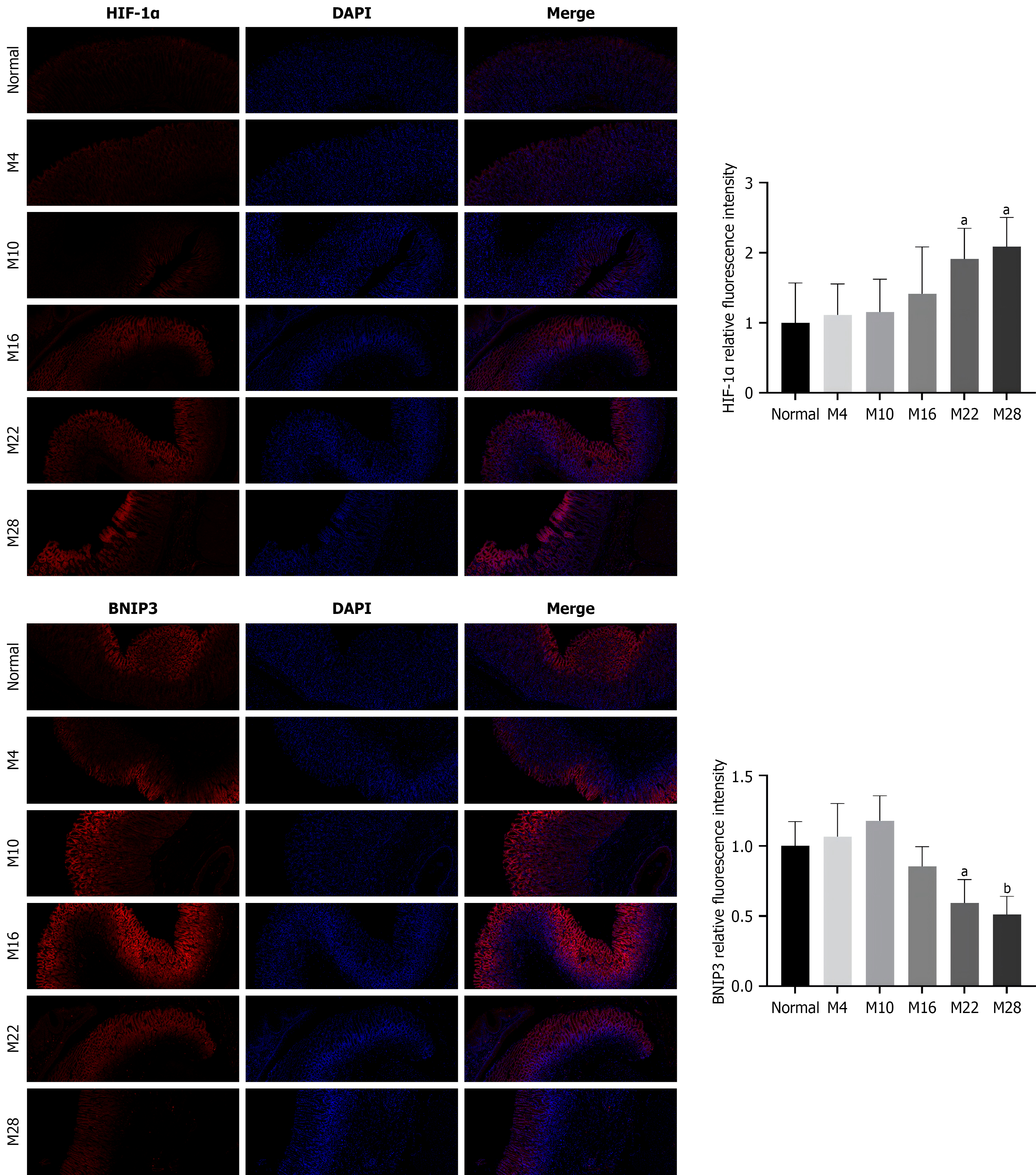
Figure 5 Representative immunofluorescence images showing the expression of hypoxia-inducible factor 1-alpha and BCL2/adenovirus E1B 19 kDa interacting protein 3 in rat gastric mucosa.
The protein expression of hypoxia-inducible factor 1-alpha and BCL2/adenovirus E1B 19 kDa interacting protein 3 was detected by immunofluorescence. Positive scores for hypoxia-inducible factor 1-alpha and BCL2/adenovirus E1B 19 kDa interacting protein 3 proteins were quantified using ImageJ software. Data are presented as the mean ± SD from six independent animals per group (n = 6). aP < 0.05, bP < 0.01 vs the normal group. BNIP3: BCL2/adenovirus E1B 19 kDa interacting protein 3; HIF-1α: Hypoxia-inducible factor 1-alpha.
- Citation: Zhang XF, Lv GH, An SJ, Yang XR, Guo WZ, Yu CY, Zhu CH. Dysregulation of the HIF-1α/BNIP3 axis links defective mitophagy to glycolytic reprogramming in a rat model of gastric precancerogenesis. World J Gastrointest Oncol 2026; 18(4): 117815
- URL: https://www.wjgnet.com/1948-5204/full/v18/i4/117815.htm
- DOI: https://dx.doi.org/10.4251/wjgo.v18.i4.117815