
















Copyright: ©Author(s) 2026.
World J Gastrointest Oncol. Apr 15, 2026; 18(4): 117815
Published online Apr 15, 2026. doi: 10.4251/wjgo.v18.i4.117815
Published online Apr 15, 2026. doi: 10.4251/wjgo.v18.i4.117815
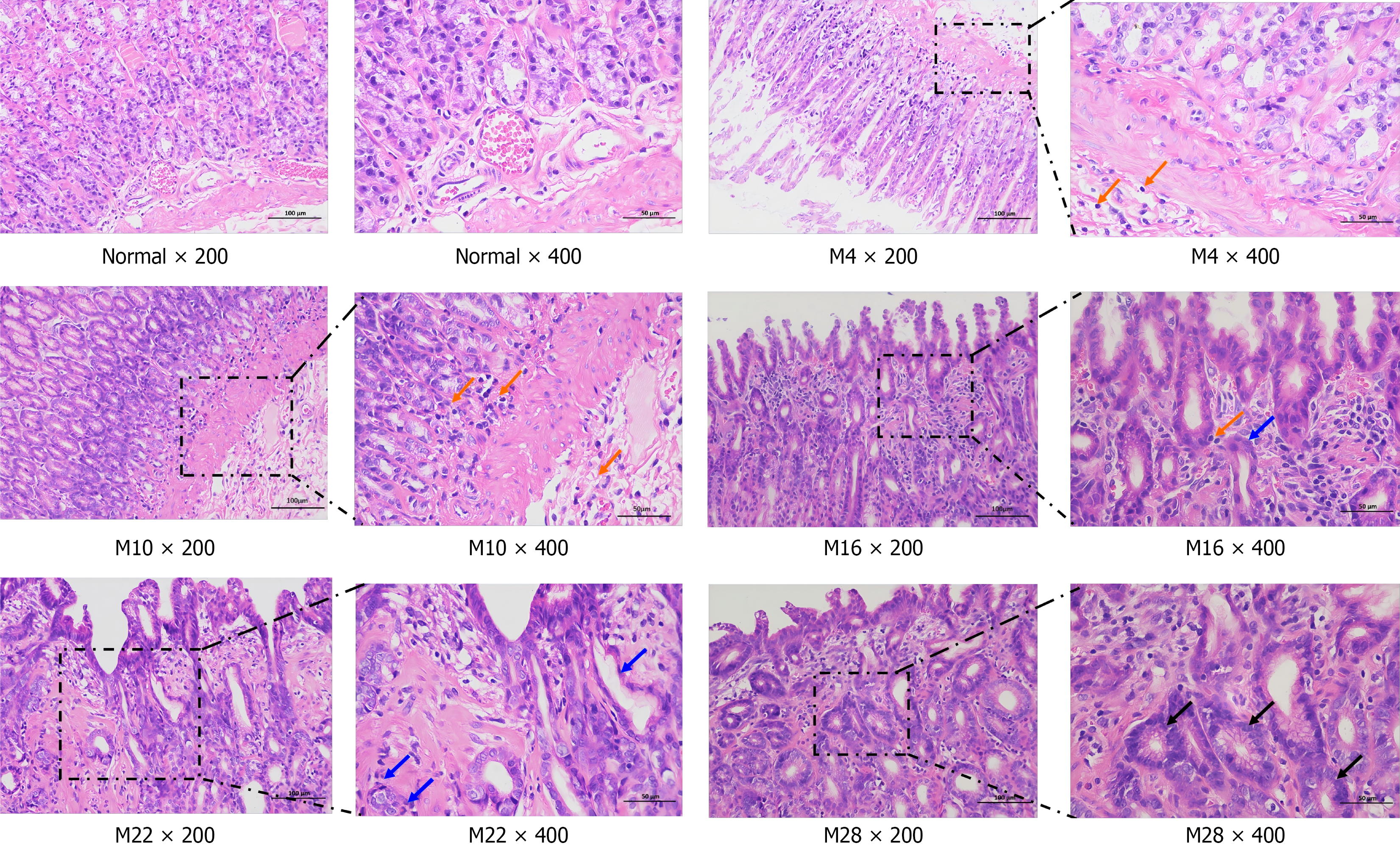
Figure 3 Histopathological changes in the rat gastric mucosa revealed by hematoxylin and eosin staining.
Representative images from the model group show progressive development of gastric lesions. At weeks 4 and 10, prominent lymphocyte infiltration was observed (orange arrow). By weeks 16 and 22, the gastric mucosa exhibited features of atrophy and intestinal metaplasia, characterized by glandular disarray, mild stromal connective tissue proliferation, and the appearance of occasional goblet cells (blue arrow). At week 28, the lesions progressed to high-grade dysplasia, featuring severe glandular disarray, nuclear hyperchromasia, and the presence of mitotic figures (black arrow). Images are shown at 200 × (scale bar = 100 μm) and 400 × (scale bar = 50 μm) magnification.
- Citation: Zhang XF, Lv GH, An SJ, Yang XR, Guo WZ, Yu CY, Zhu CH. Dysregulation of the HIF-1α/BNIP3 axis links defective mitophagy to glycolytic reprogramming in a rat model of gastric precancerogenesis. World J Gastrointest Oncol 2026; 18(4): 117815
- URL: https://www.wjgnet.com/1948-5204/full/v18/i4/117815.htm
- DOI: https://dx.doi.org/10.4251/wjgo.v18.i4.117815