
















Copyright: ©Author(s) 2026.
World J Gastrointest Oncol. Apr 15, 2026; 18(4): 116104
Published online Apr 15, 2026. doi: 10.4251/wjgo.v18.i4.116104
Published online Apr 15, 2026. doi: 10.4251/wjgo.v18.i4.116104
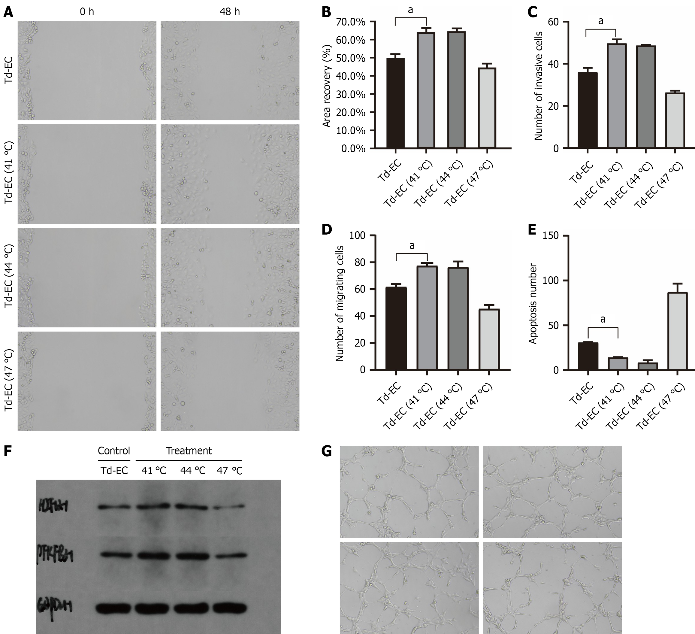
Figure 3 Functional changes in tumor-derived endothelial cells after thermal stimulation.
A: Wound-healing migration assay at 0 h and 48 h under 37 °C or 41 °C (n = 3); B: Quantification of wound-closure rate (n = 3); C: Transwell migration assay (n = 3); D: Transwell invasion assay (n = 3); E: Apoptosis measured by TUNEL staining at 37 °C, 41 °C, 44 °C, and 47 °C (n = 3); F: Western blot analysis of hypoxia-inducible factor-1α, 6-phosphofructo-2-kinase/fructose-2,6-bisphosphatase 3, and GAPDH under each temperature condition (n = 3); G: Tube-formation assay under 37 °C, 41 °C, 44 °C, and 47 °C (n = 3). Data are expressed as mean ± SD; one-way ANOVA was used for statistical analysis. aP < 0.05. Td-EC: Tumor-derived endothelial cells.
- Citation: Lu D, Wang LJ, Chai J, Jiang J, Tang YL. MicroRNA-206 suppresses hypoxia-inducible factor-1α/PFKFB3-mediated glycolysis to inhibit recurrence and metastasis of hepatocellular carcinoma after incomplete radiofrequency ablation. World J Gastrointest Oncol 2026; 18(4): 116104
- URL: https://www.wjgnet.com/1948-5204/full/v18/i4/116104.htm
- DOI: https://dx.doi.org/10.4251/wjgo.v18.i4.116104