
















Copyright: ©Author(s) 2026.
World J Gastrointest Oncol. Apr 15, 2026; 18(4): 116097
Published online Apr 15, 2026. doi: 10.4251/wjgo.v18.i4.116097
Published online Apr 15, 2026. doi: 10.4251/wjgo.v18.i4.116097
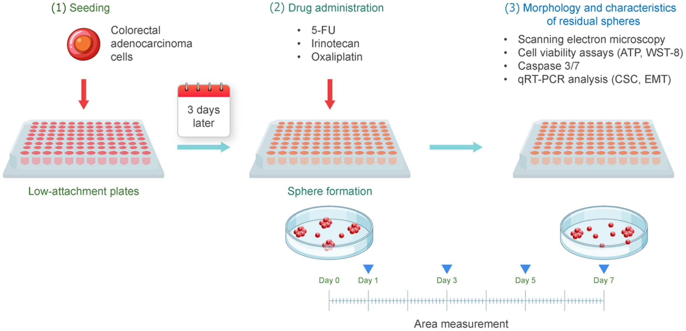
Figure 1 Schema of the experimental method.
Colorectal adenocarcinoma cells were cultured in low-attachment 96-well plates for 3 days to form spheres. After sphere formation, anticancer drugs were added to each well at a concentration of 30 μM. The day of anticancer drug administration was defined as day 0, and the areas of the spheres were measured on days 0, 1, 3, 5, and 7 using an imaging system. 5-FU: 5-fluorouracil; WST-8: Water-soluble tetrazolium 8; qRT-PCR: Quantitative reverse transcriptase PCR; CSC: Cancer stem cell; EMT: Epithelial-mesenchymal transition.
- Citation: Yoshimura Y, Shinji S, Ogawa Y, Yamada T, Matsuda A, Uehara K, Yokoyama Y, Takahashi G, Iwai T, Miyasaka T, Kanaka S, Matsui T, Hayashi K, Fujiwara M, Shichi Y, Arai T, Ishiwata T, Yoshida H. Morphological and functional responses of three-dimensional-cultured colorectal cancer spheres to anticancer drugs. World J Gastrointest Oncol 2026; 18(4): 116097
- URL: https://www.wjgnet.com/1948-5204/full/v18/i4/116097.htm
- DOI: https://dx.doi.org/10.4251/wjgo.v18.i4.116097