
















Copyright: ©Author(s) 2026.
World J Gastrointest Oncol. Apr 15, 2026; 18(4): 114547
Published online Apr 15, 2026. doi: 10.4251/wjgo.v18.i4.114547
Published online Apr 15, 2026. doi: 10.4251/wjgo.v18.i4.114547
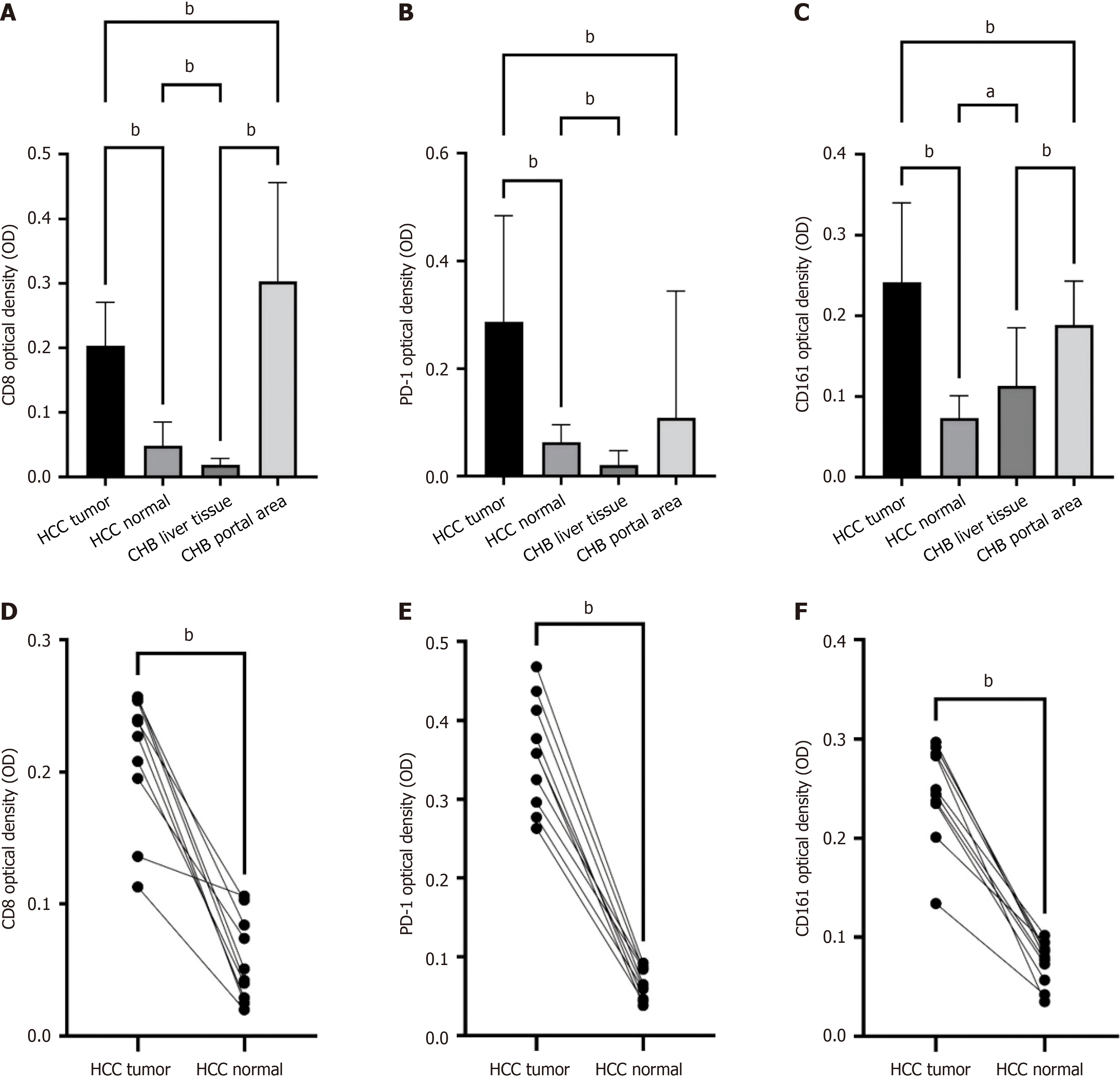
Figure 8 Semiquantitative unpaired analysis of CD161, CD8, and programmed death receptor 1 immunohistochemical results.
A: CD8 optical density (OD) of hepatocellular carcinoma (HCC) normal tissues, HCC tumor tissues, chronic hepatitis B (CHB) liver tissues and CHB portal area tissues; B: Programmed death receptor 1 (PD-1) OD of HCC normal tissues, HCC tumor tissues, CHB liver tissues and CHB portal area tissues; C: CD161 OD of HCC normal tissues, HCC tumor tissues, CHB liver tissues and CHB portal area tissues; D: Paired analysis of CD8; E: Paired analysis of PD-1; F: Paired analysis of CD161. A-C: ANOVA; D-F: Paired t test. aP < 0.05; bP < 0.01. PD-1: Programmed death receptor 1; CHB: Chronic hepatitis B; HCC: Hepatocellular carcinoma; OD: Optical density.
- Citation: Liu CR, Zhang M, Wang MQ, Zhang W, Li J, Shi GZ, Liang FF, Li YP, Huang N. CD161+CD8+ T cells in patients with hepatitis B virus-associated hepatocellular carcinoma. World J Gastrointest Oncol 2026; 18(4): 114547
- URL: https://www.wjgnet.com/1948-5204/full/v18/i4/114547.htm
- DOI: https://dx.doi.org/10.4251/wjgo.v18.i4.114547