
















Copyright: ©Author(s) 2026.
World J Gastrointest Oncol. Mar 15, 2026; 18(3): 116436
Published online Mar 15, 2026. doi: 10.4251/wjgo.v18.i3.116436
Published online Mar 15, 2026. doi: 10.4251/wjgo.v18.i3.116436
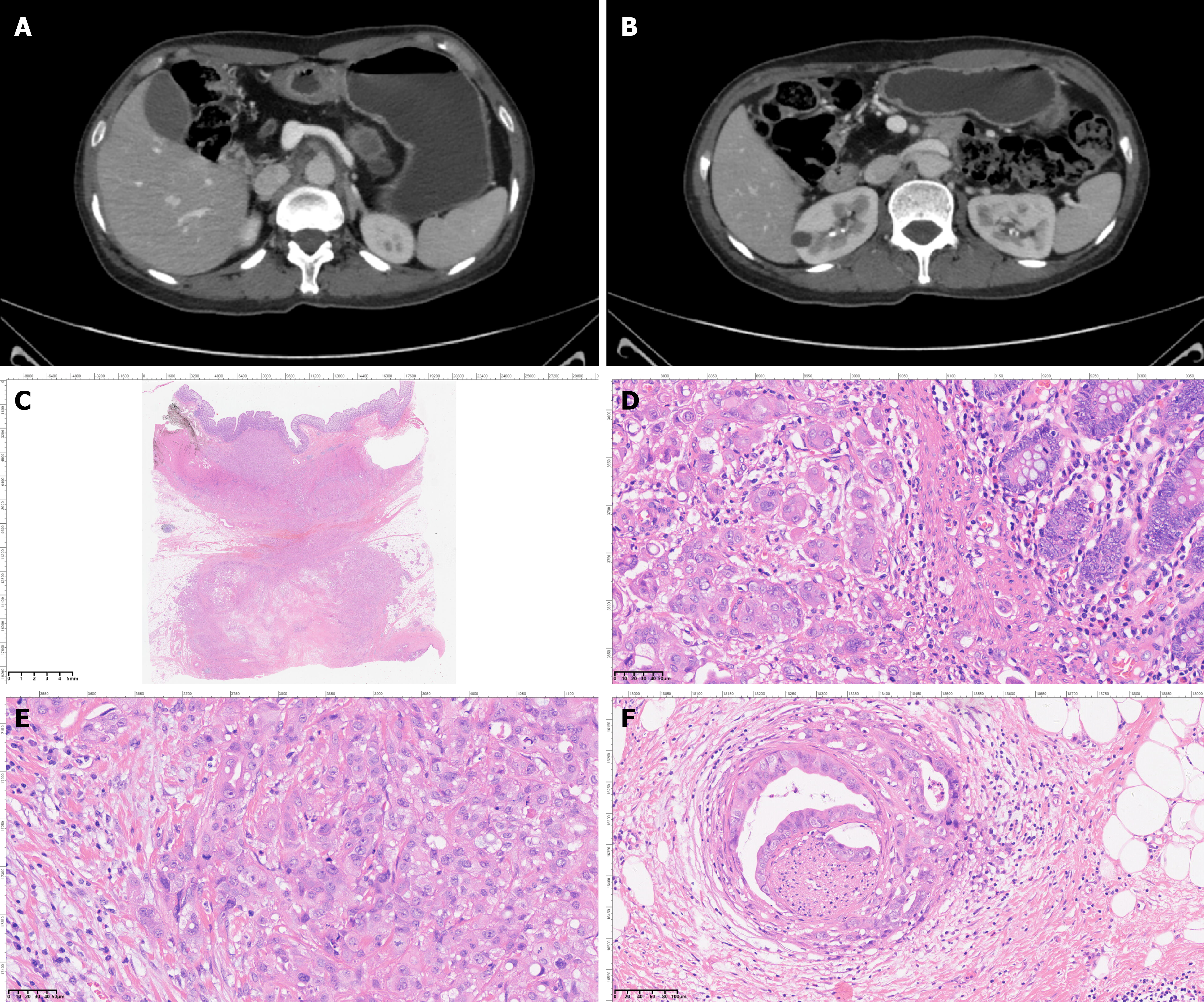
Figure 3 Upper abdomen contrast-enhanced dynamic computed tomography and histopathological analysis of the resected specimen.
A and B: Pancreatic parenchymal atrophy, multiple calcifications in the pancreatic head and body, and a non-enhancing hypodense lesion in the pancreatic tail are observed; C: Malignant tumor cells were observed in the pathological specimen of pancreatic body and tail resection combined with partial colectomy; D: Tumor cell infiltration was observed in the colon; E: A large amount of ductal adenocarcinoma was observed in the pancreas; F: Perineural invasion by carcinoma was observed in the surrounding stroma.
- Citation: Tao ZT, Xu L, Chen YL, Gong B. Pancreatic ductal adenocarcinoma developing from hereditary pancreatitis: A case report. World J Gastrointest Oncol 2026; 18(3): 116436
- URL: https://www.wjgnet.com/1948-5204/full/v18/i3/116436.htm
- DOI: https://dx.doi.org/10.4251/wjgo.v18.i3.116436