
















Copyright: ©Author(s) 2026.
World J Gastrointest Oncol. Mar 15, 2026; 18(3): 115905
Published online Mar 15, 2026. doi: 10.4251/wjgo.v18.i3.115905
Published online Mar 15, 2026. doi: 10.4251/wjgo.v18.i3.115905
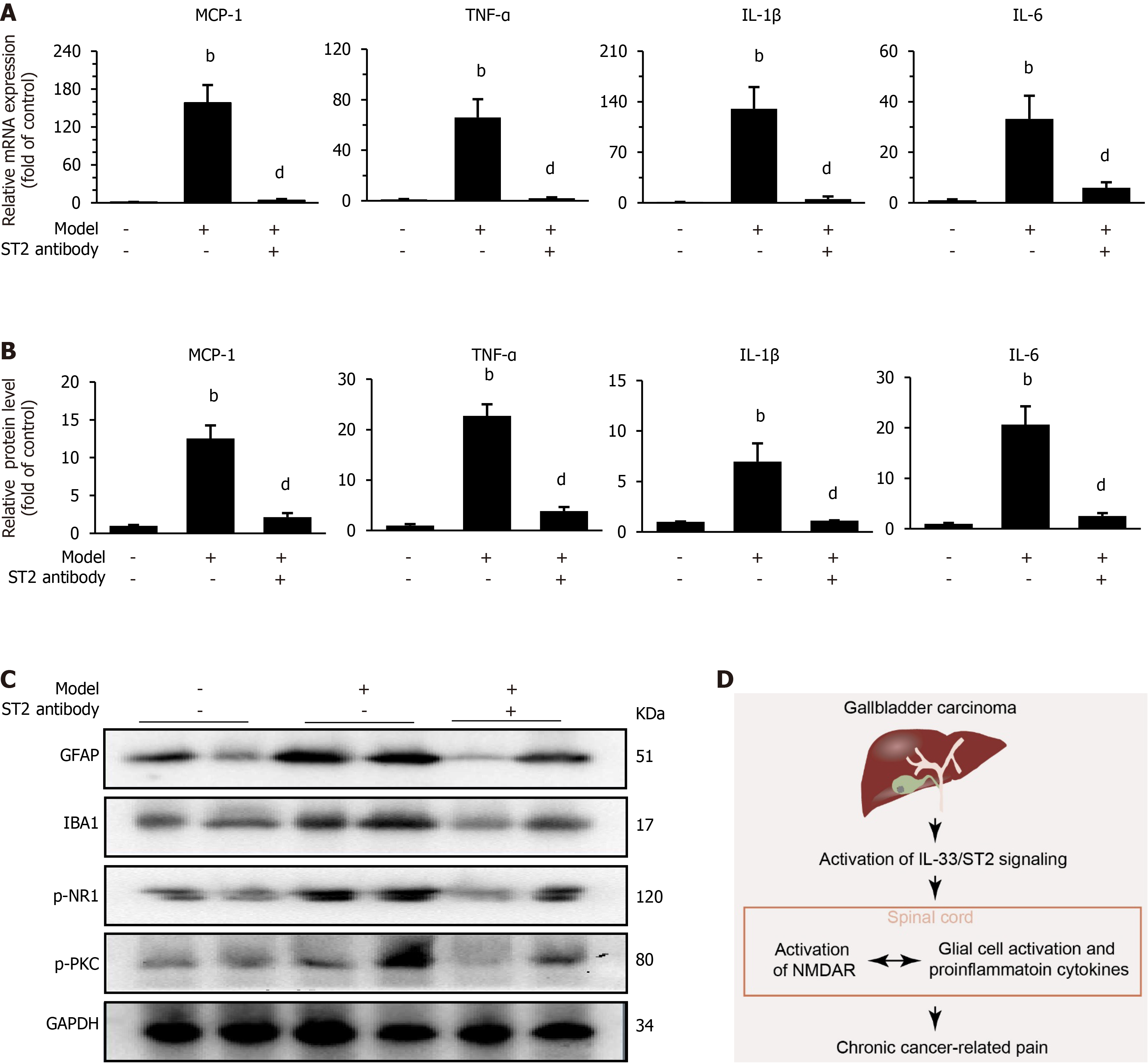
Figure 4 Intrathecal administration of the anti-suppression of tumorigenicity 2 antibody suppresses glial cell activation, proinflammatory cytokine release and phosphorylation of the N-methyl-D-aspartate receptor subunit 1 and protein kinase C in the spinal cords of animals with gallbladder carcinoma-induced chronic pain.
A and B: MRNA and protein levels of monocyte chemoattractant protein 1, tumour necrosis factor-α, interleukin (IL)-1β, and IL-6 in the spinal cords of model mice with gallbladder carcinoma-induced chronic pain; C: Western blot analysis showing the effects of repeated intrathecal injections of the anti-suppression of tumorigenicity 2 antibody (300 ng, daily for three consecutive days 50-52 days after the operation) on the expression of glial fibrillary acidic protein, ionized calcium-binding adaptor molecule 1, p-N-methyl-D-aspartate receptor subunit 1, and p-protein kinase C; D: Proposed mechanisms of action by which IL-33/suppression of tumorigenicity 2 signaling promotes gallbladder carcinoma-induced chronic pain. Tissues were collected on the 52nd day, 2 hours after the last injection. Six spinal cord segments were included in each of the groups. One-way ANOVA. bP < 0.01 vs control, and dP < 0.01 vs model + saline. ST2: Suppression of tumorigenicity 2; MCP-1: Monocyte chemoattractant protein 1; TNF: Tumour necrosis factor; IL: Interleukin; GFAP: Glial fibrillary acidic protein; IBA1: Ionized calcium-binding adaptor molecule 1; NR1: N-methyl-D-aspartate receptor subunit 1; PKC: Protein kinase C; GAPDH: Glyceraldehyde 3-phosphate dehydrogenase; NMDAR: N-methyl-D-aspartic acid receptor.
- Citation: Ge XX, Dong XM, Yang Y, Zhang W, Zhou W, Deng XT. Activation of interleukin-33/suppression of tumorigenicity 2 signaling contributes to gallbladder carcinoma-induced chronic pain. World J Gastrointest Oncol 2026; 18(3): 115905
- URL: https://www.wjgnet.com/1948-5204/full/v18/i3/115905.htm
- DOI: https://dx.doi.org/10.4251/wjgo.v18.i3.115905