
















Copyright: ©Author(s) 2026.
World J Gastrointest Oncol. Mar 15, 2026; 18(3): 115812
Published online Mar 15, 2026. doi: 10.4251/wjgo.v18.i3.115812
Published online Mar 15, 2026. doi: 10.4251/wjgo.v18.i3.115812
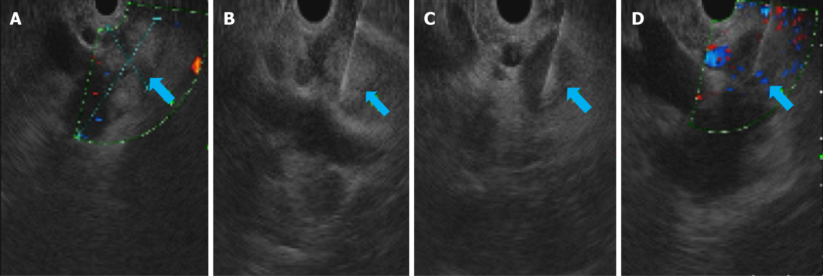
Figure 2 Endoscopic ultrasound-guided fine-needle aspiration biopsy procedure.
A: Ultrasound image of the liver (S1 segment) showing a lesion characterized by low-level echoes, measuring approximately 50.5 mm × 25.5 mm (arrow). Internal changes, including high-intensity echoes, were observed within the lesion. It was observed that the lesion surrounded the portal vein and its confluence and was in proximity to the lower segment of the hepatic portal vein. The surrounding low-intensity lymph nodes appear dispersed; B and C: Ultrasound-guided puncture at the lesion site (arrow); D: Perifocal lymph node puncture (arrow).
- Citation: Qi DY, Zhao YC, Chen LP. Isolated hepatic tuberculoma: A case report and review of the literature. World J Gastrointest Oncol 2026; 18(3): 115812
- URL: https://www.wjgnet.com/1948-5204/full/v18/i3/115812.htm
- DOI: https://dx.doi.org/10.4251/wjgo.v18.i3.115812