
















Copyright: ©Author(s) 2026.
World J Gastrointest Oncol. Mar 15, 2026; 18(3): 115737
Published online Mar 15, 2026. doi: 10.4251/wjgo.v18.i3.115737
Published online Mar 15, 2026. doi: 10.4251/wjgo.v18.i3.115737
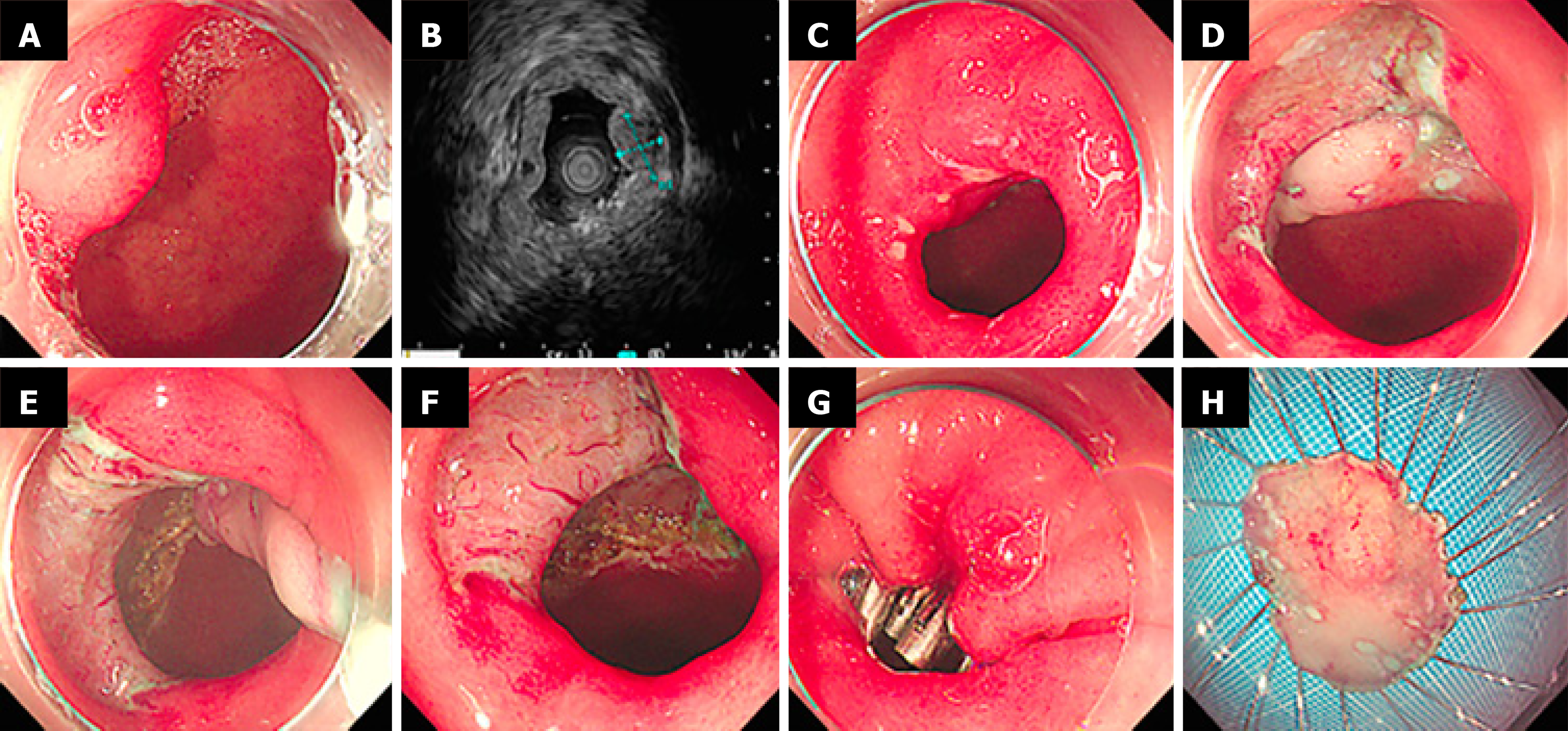
Figure 3 Endoscopic submucosal dissection for sporadic nonampullary duodenal tumor.
Submucosal tumor, 10 mm × 6 mm, neuroendocrine tumor G1, vertical margin (-), horizontal margin (-). A: Lesion in the bulb; B: Micro-probe endoscopic ultrasonography showed a hypoechoic lesion in the second layer; C: Marking; D and E: Dissection with golden knife; F: Post-endoscopic submucosal dissection ulcer; G: Clip closure; H: En bloc specimen.
- Citation: Yu H, Rong L, Cai YL, Liu GY, Guo XY. Short- and long-term outcomes after endoscopic resection of sporadic nonampullary duodenal tumors: An 11-year single-center retrospective study. World J Gastrointest Oncol 2026; 18(3): 115737
- URL: https://www.wjgnet.com/1948-5204/full/v18/i3/115737.htm
- DOI: https://dx.doi.org/10.4251/wjgo.v18.i3.115737