
















Copyright: ©Author(s) 2026.
World J Gastrointest Oncol. Mar 15, 2026; 18(3): 113851
Published online Mar 15, 2026. doi: 10.4251/wjgo.v18.i3.113851
Published online Mar 15, 2026. doi: 10.4251/wjgo.v18.i3.113851
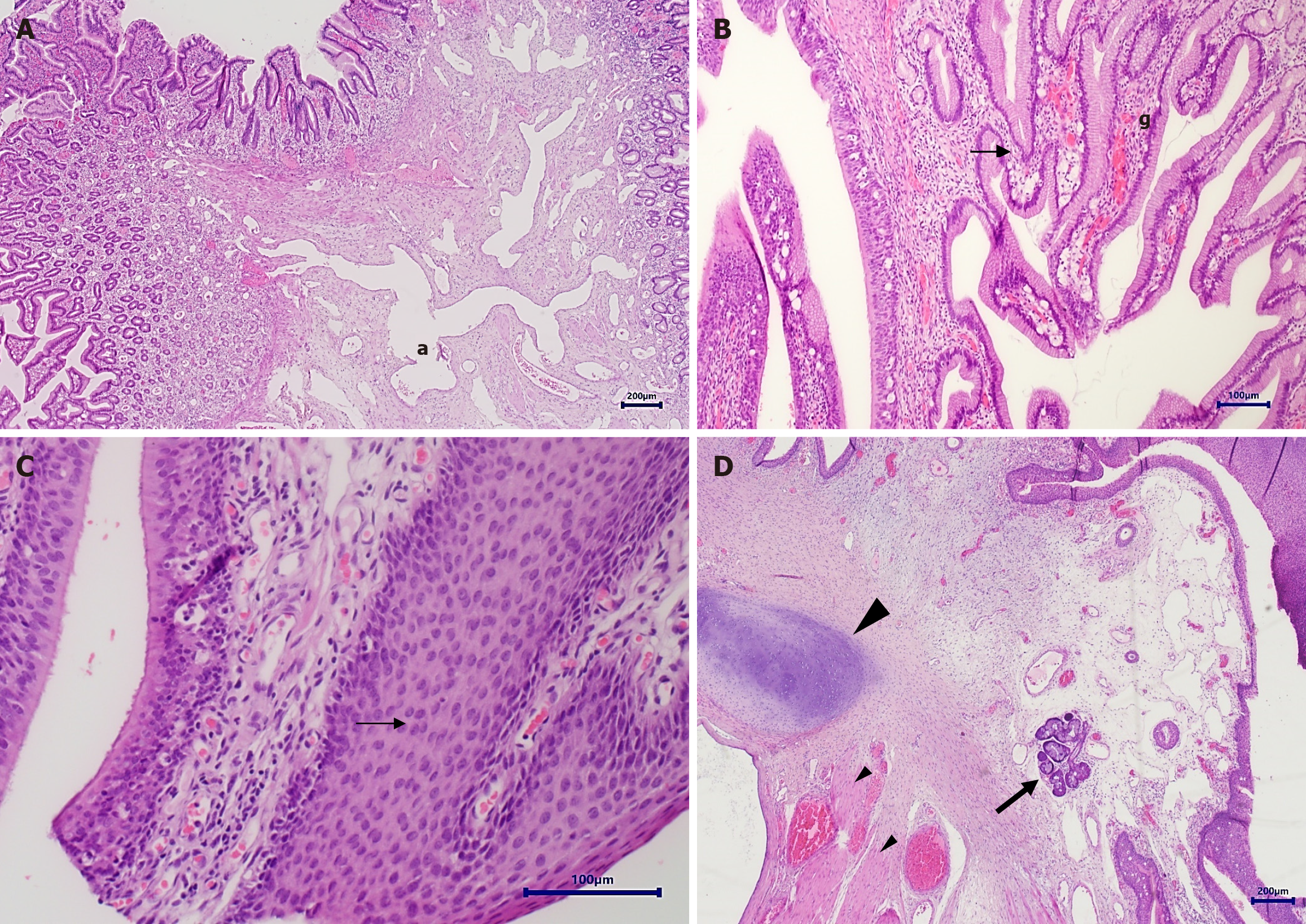
Figure 5 Pathology slices of the tumour.
Hematoxylin and eosin-stained sections from esophageal choristoma. A: Sections show a mass lined mainly by gastric mucosa (left upper and right), with submucosal edema and dilated vascular channels (a); B: Respiratory type ciliated epithelium (arrow) is adjacent to gastric mucosa (g); C: Squamous epithelium is present (arrow); D: Additional features include cartilage plates consistent with tracheobronchial remnants (large arrowhead), and occasional salivary glands (arrow).
- Citation: Giz N, Epshtein J, Savin B, Meir K, Slae M. Presentation and endoscopic resection technique of neonatal esophageal choristoma: A case report. World J Gastrointest Oncol 2026; 18(3): 113851
- URL: https://www.wjgnet.com/1948-5204/full/v18/i3/113851.htm
- DOI: https://dx.doi.org/10.4251/wjgo.v18.i3.113851