
















Copyright: ©Author(s) 2026.
World J Gastrointest Oncol. Mar 15, 2026; 18(3): 113851
Published online Mar 15, 2026. doi: 10.4251/wjgo.v18.i3.113851
Published online Mar 15, 2026. doi: 10.4251/wjgo.v18.i3.113851
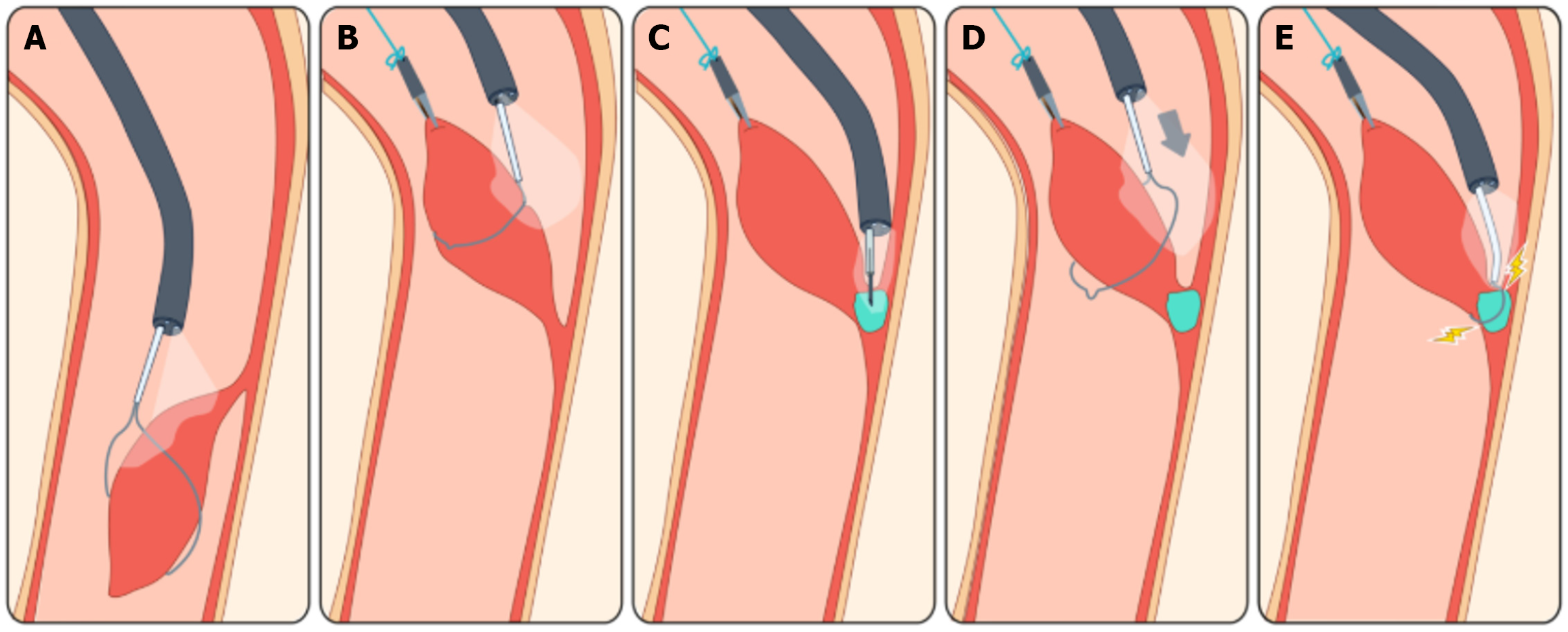
Figure 4 Illustration of the procedure.
A: A slim endoscope was advanced down the esophagus along the longitudinal axis of the mass, which was oriented caudally. A slim-scope compatible snare was used to flip the mass cephalad; B: A hemoclip was applied under direct vision to the tip of the mass. The hemoclip secured a suture wire, allowing upward traction to be exerted on the mass; C: An injection needle sized for the slim scope was used to inject the base of the lesion with an adrenaline-saline solution (1 mg adrenaline in 10 mL normal saline, total volume 2 mL) to create a submucosal lift; D: A slim-scope compatible snare was then inserted through the endoscope and advanced over the traction wire, the clip, and the mass body until it reached the lesion’s base; E: Resection of the mass was performed using a hot-polypectomy technique (ERBE VIO 300 Endocut Q, effect 2, cut duration 1, cut interval 6, maximum output 770).
- Citation: Giz N, Epshtein J, Savin B, Meir K, Slae M. Presentation and endoscopic resection technique of neonatal esophageal choristoma: A case report. World J Gastrointest Oncol 2026; 18(3): 113851
- URL: https://www.wjgnet.com/1948-5204/full/v18/i3/113851.htm
- DOI: https://dx.doi.org/10.4251/wjgo.v18.i3.113851