
















©The Author(s) 2026.
World J Gastrointest Oncol. Feb 15, 2026; 18(2): 115689
Published online Feb 15, 2026. doi: 10.4251/wjgo.v18.i2.115689
Published online Feb 15, 2026. doi: 10.4251/wjgo.v18.i2.115689
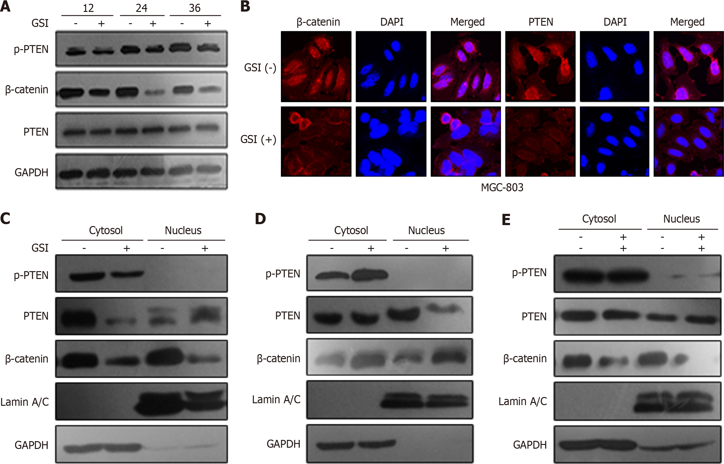
Figure 3 Presenilin-1 regulated tensin homolog deleted on chromosome ten phosphorylation and subcellular localization through activating β-catenin.
A: MGC-803 cells were treated with 1 μM γ-secretase inhibitor (GSI) for 12 h, 24 h, and 36 h. Time-dependent protein expressions of β-catenin, phosphorylation of tensin homolog deleted on chromosome ten (PTEN), and PTEN with or without GSI treatment were detected via western blot analysis. Glyceraldehyde-3-phosphate dehydrogenase was used as a loading control; B: Localization of β-catenin and PTEN after GSI treatment. Cells were treated with 1 μM GSI for 24 h, and immunocytochemical analysis was performed with different antibodies. Nuclei were detected by DAPI staining; C-E: Subcellular localization of phosphorylation of PTEN, PTEN, and β-catenin after treatment of GSI, lentivirus presenilin-1, and both lentivirus presenilin-1 and β-catenin small interfering RNA. Cytoplasmic and nuclear fractions were isolated via the subcellular fraction assay. Glyceraldehyde-3-phosphate dehydrogenase was used as a control in the cytoplasm and lamin A/C in the nucleus. GSI: Γ-secretase inhibitor; p-PTEN: Phosphorylation of tensin homolog deleted on chromosome ten; PTEN: Tensin homolog deleted on chromosome ten; GAPDH: Glyceraldehyde-3-phosphate dehydrogenase.
- Citation: Lin X, Lin GF, Gu FT, Li YL. Increasing expression of presenilin 1, β-catenin, and p-PTEN and its regulatory roles on cell invasion in gastric cancer. World J Gastrointest Oncol 2026; 18(2): 115689
- URL: https://www.wjgnet.com/1948-5204/full/v18/i2/115689.htm
- DOI: https://dx.doi.org/10.4251/wjgo.v18.i2.115689