
















©The Author(s) 2026.
World J Gastrointest Oncol. Feb 15, 2026; 18(2): 113494
Published online Feb 15, 2026. doi: 10.4251/wjgo.v18.i2.113494
Published online Feb 15, 2026. doi: 10.4251/wjgo.v18.i2.113494
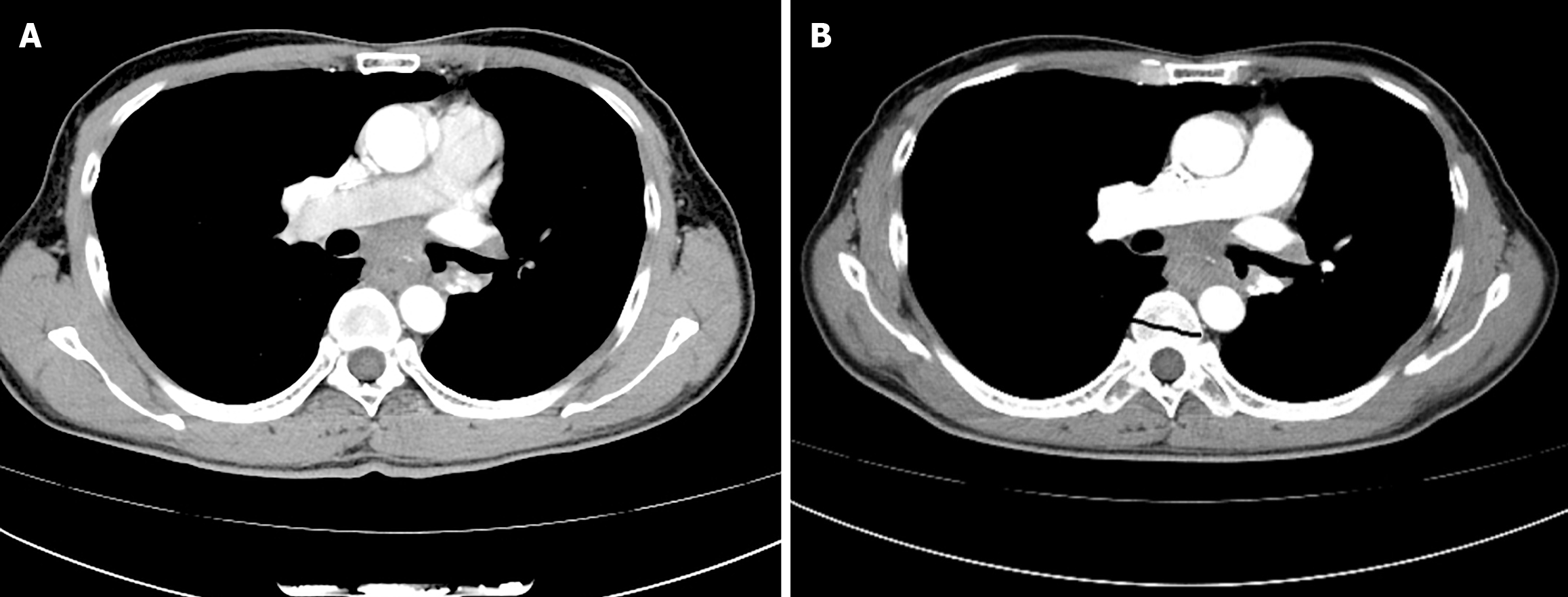
Figure 3 Chest computed tomography imaging.
A: Showing mid-esophageal wall thickening and subcarinal lymphadenopathy when the patient presented to the thoracic surgeon; B: Showing the volume of the esophageal tumor increased, causing the esophageal lumen to narrow, after two cycles of neoadjuvant immunochemotherapy.
- Citation: Zhang Y, Li ZX, Ma DY, Liu F. Solitary esophageal metastasis ten years after curative resection of stage I rectal adenocarcinoma: A case report. World J Gastrointest Oncol 2026; 18(2): 113494
- URL: https://www.wjgnet.com/1948-5204/full/v18/i2/113494.htm
- DOI: https://dx.doi.org/10.4251/wjgo.v18.i2.113494