
















Copyright: ©Author(s) 2026.
World J Gastrointest Oncol. Nov 15, 2026; 18(11): 124110
Published online Nov 15, 2026. doi: 10.4251/wjgo.124110
Published online Nov 15, 2026. doi: 10.4251/wjgo.124110
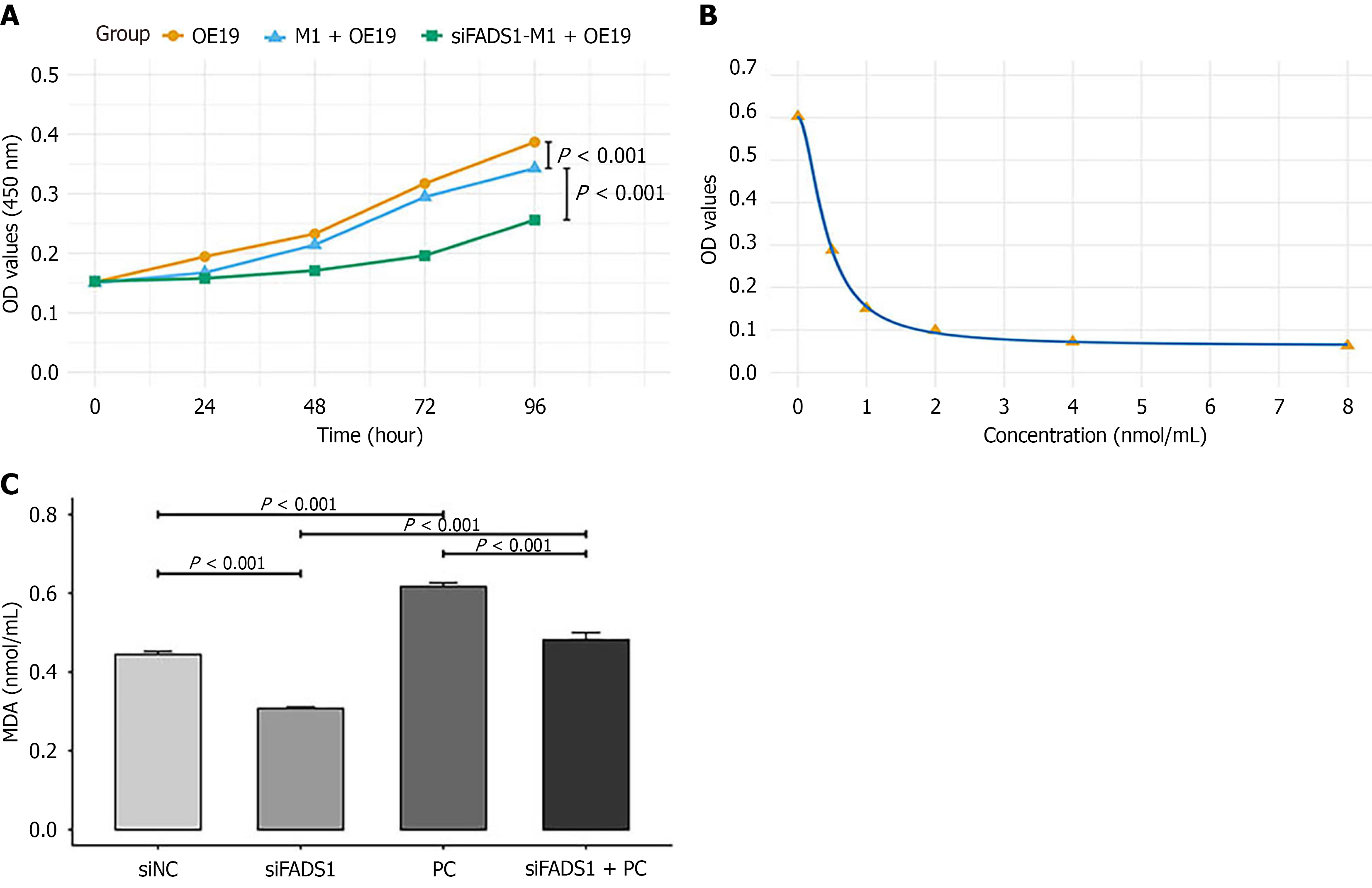
Figure 6 Fatty acid desaturase 1 promotes M1 macrophage ferroptosis via 1-palmitoyl-2-stearoyl-glycerophosphocholine (16:0/18:0).
A: OE-19 cells were cultured alone or cocultured with M1 macrophages transfected with siRNA negative control or si-fatty acid desaturase 1-1 in a transwell system for 96 hours. Cell viability was assessed by a cell counting kit-8 assay at the indicated time points and expressed as optical density at 450 nm values. The data are shown as the mean ± SD of three independent experiments (n = 6 replicates per group); B and C: M1 macrophages were transfected with siRNA negative control or si-fatty acid desaturase 1-1 and treated with or without 10 μM phosphatidylcholine (16:0/18:0) for 24 hours, after which the malondialdehyde concentration was measured. The data are presented as the mean ± SD from three independent experiments. FADS: Fatty acid desaturase; MDA: Malon
- Citation: Du QC, Bai Y, Zhou DH, Yu H, Nian H. Fatty acid desaturase 1 drives esophageal cancer via M1 macrophage ferroptosis and lipid metabolism remodeling. World J Gastrointest Oncol 2026; 18(11): 124110
- URL: https://www.wjgnet.com/1948-5204/full/v18/i11/124110.htm
- DOI: https://dx.doi.org/10.4251/wjgo.124110