
















Copyright: ©Author(s) 2026.
World J Gastrointest Oncol. Oct 15, 2026; 18(10): 124211
Published online Oct 15, 2026. doi: 10.4251/wjgo.124211
Published online Oct 15, 2026. doi: 10.4251/wjgo.124211
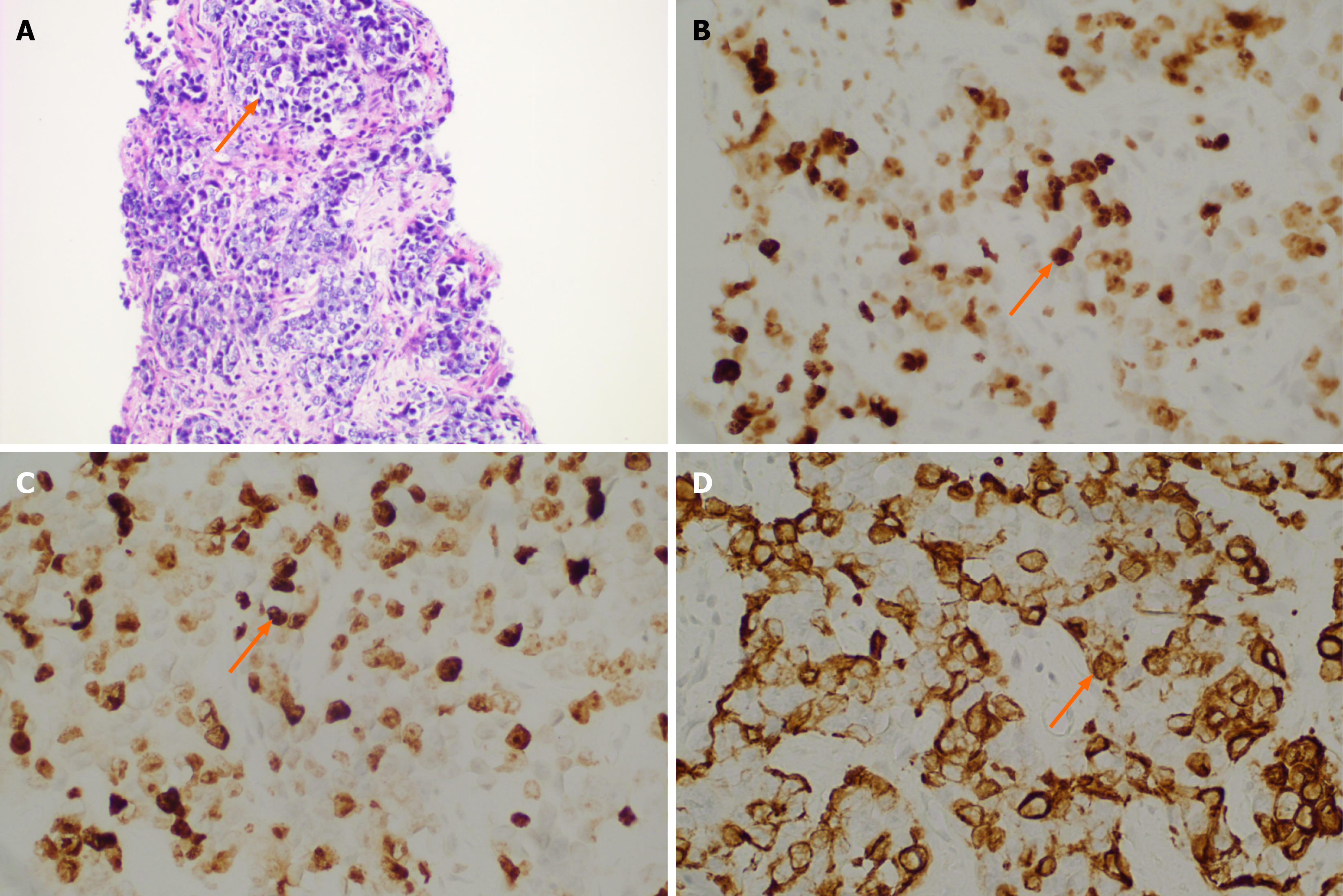
Figure 2 Histopathological and immunohistochemical features of the liver biopsy specimen.
All hematoxylin and eosin (H&E) and immunohistochemical panels were obtained from the same biopsy specimen. A: H&E staining shows abundant tumor cells (original magnification, × 200); B: Immunohistochemistry shows diffuse nuclear positivity for p63 (original magnification, × 400); C: Nuclear positivity for p40 (original magnification, × 400); D: Cytoplasmic positivity for cytokeratin 5/6 (CK5/6; original magnification, × 400). The combined expression of p63, p40, and CK5/6 supports squamous differentiation of the tumor.
- Citation: Qing D, Pan SL, Zhao J, Liu XB, Li WN. Primary squamous cell carcinoma arising in the liver: A case report and review of literature. World J Gastrointest Oncol 2026; 18(10): 124211
- URL: https://www.wjgnet.com/1948-5204/full/v18/i10/124211.htm
- DOI: https://dx.doi.org/10.4251/wjgo.124211