
















Copyright: ©Author(s) 2026.
World J Gastrointest Oncol. Oct 15, 2026; 18(10): 123703
Published online Oct 15, 2026. doi: 10.4251/wjgo.123703
Published online Oct 15, 2026. doi: 10.4251/wjgo.123703
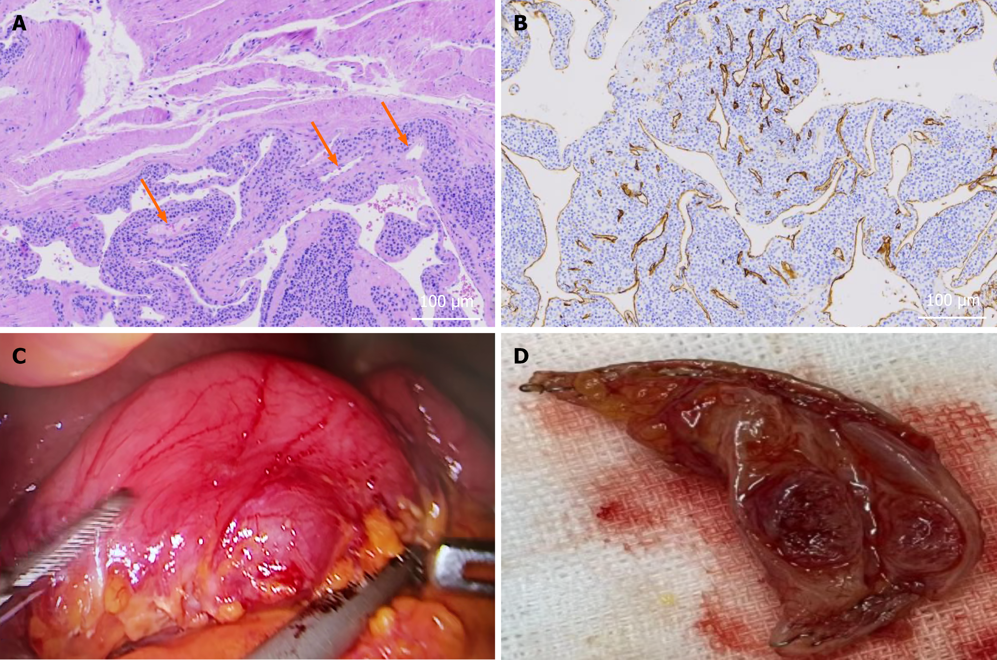
Figure 2 Surgical and pathological findings of the gastric glomus tumor.
A: Hematoxylin and eosin staining showed nests and sheets of relatively uniform round tumor cells arranged around dilated vascular channels (arrows; scale bar: 100 μm); B: Immunohistochemical staining demonstrated positive expression of smooth muscle actin in the tumor cells; C: Intraoperative laparoscopic view revealed a protruding mass on the gastric wall; D: Gross appearance of the resected specimen showed a well-circumscribed nodular lesion.
- Citation: Luo YK, Yu HY, Bao YP, Mao JQ, Chen YB. Double contrast-enhanced ultrasonography in an exophytic gastric glomus tumor with unremarkable gastroscopy: A case report. World J Gastrointest Oncol 2026; 18(10): 123703
- URL: https://www.wjgnet.com/1948-5204/full/v18/i10/123703.htm
- DOI: https://dx.doi.org/10.4251/wjgo.123703