
















Copyright: ©Author(s) 2026.
World J Gastrointest Oncol. Oct 15, 2026; 18(10): 123447
Published online Oct 15, 2026. doi: 10.4251/wjgo.123447
Published online Oct 15, 2026. doi: 10.4251/wjgo.123447
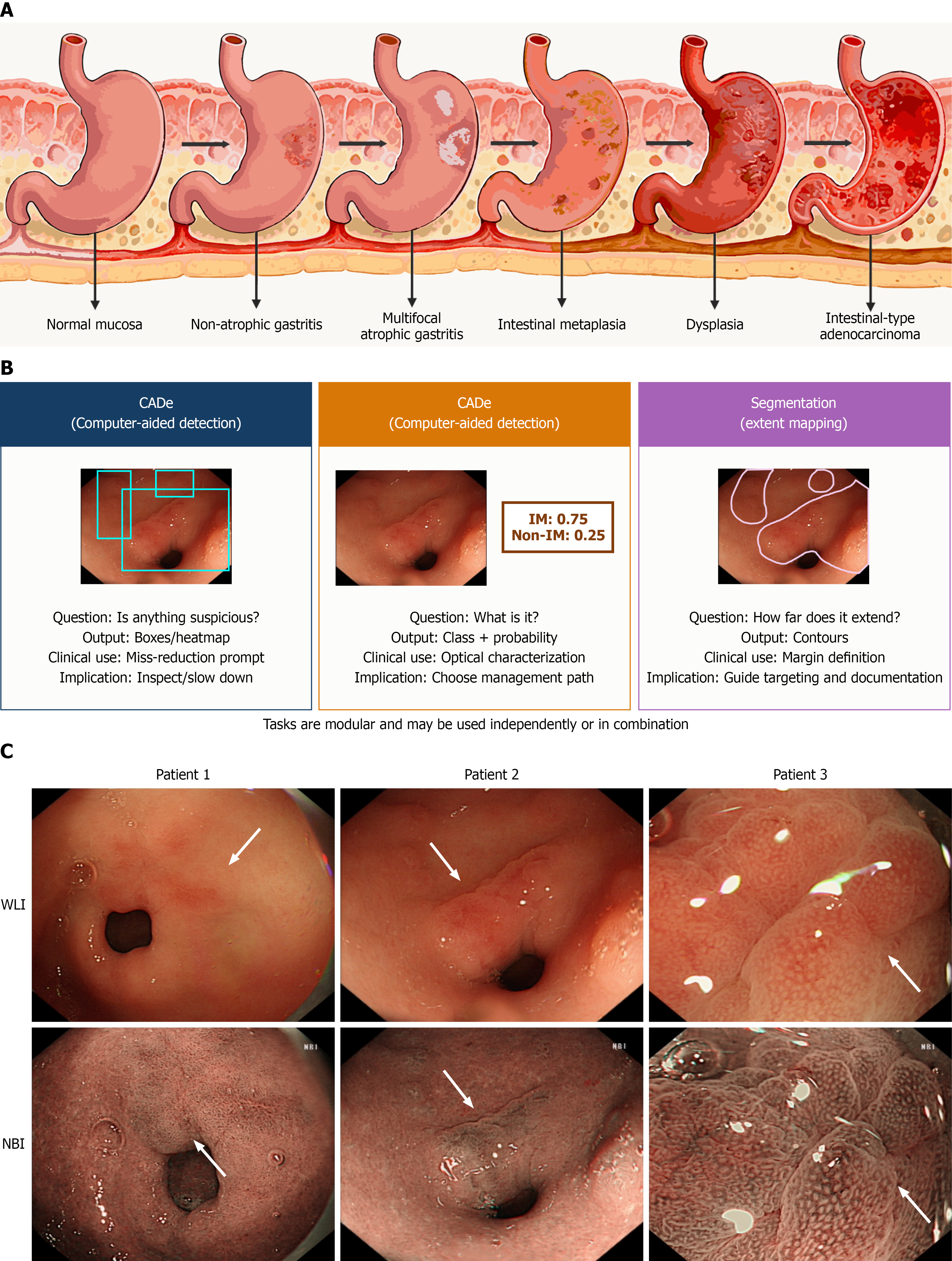
Figure 1 Overview of gastric carcinogenesis and artificial intelligence-assisted endoscopic imaging.
A: Schematic illustration of Correa’s cascade; B: Modular endoscopic artificial intelligence tasks, including computer-aided detection, computer-aided diagnosis, and segmentation (extent mapping), which may be used independently or in combination; C: Representative anonymized endoscopic images from three different patients, with each row showing paired white-light imaging and narrow-band imaging images from the same patient. The left column shows white-light imaging, and the right column shows narrow-band imaging. Arrows indicate corresponding subtle mucosal or vascular abnormalities in the paired images, which are generally more conspicuous with image-enhanced endoscopy. IM: Intestinal metaplasia; WLI: White-light imaging; NBI: Narrow-band imaging; CADe: Computer-aided detection.
- Citation: Yu HH, Chan IN, Wang JH, Qin YY, Chan IW, Wong PK. Artificial intelligence for endoscopic correlates of Correa’s cascade in gastric precancerous lesions and early neoplasia. World J Gastrointest Oncol 2026; 18(10): 123447
- URL: https://www.wjgnet.com/1948-5204/full/v18/i10/123447.htm
- DOI: https://dx.doi.org/10.4251/wjgo.123447