
















Copyright: ©Author(s) 2026.
World J Gastrointest Oncol. Oct 15, 2026; 18(10): 119879
Published online Oct 15, 2026. doi: 10.4251/wjgo.119879
Published online Oct 15, 2026. doi: 10.4251/wjgo.119879
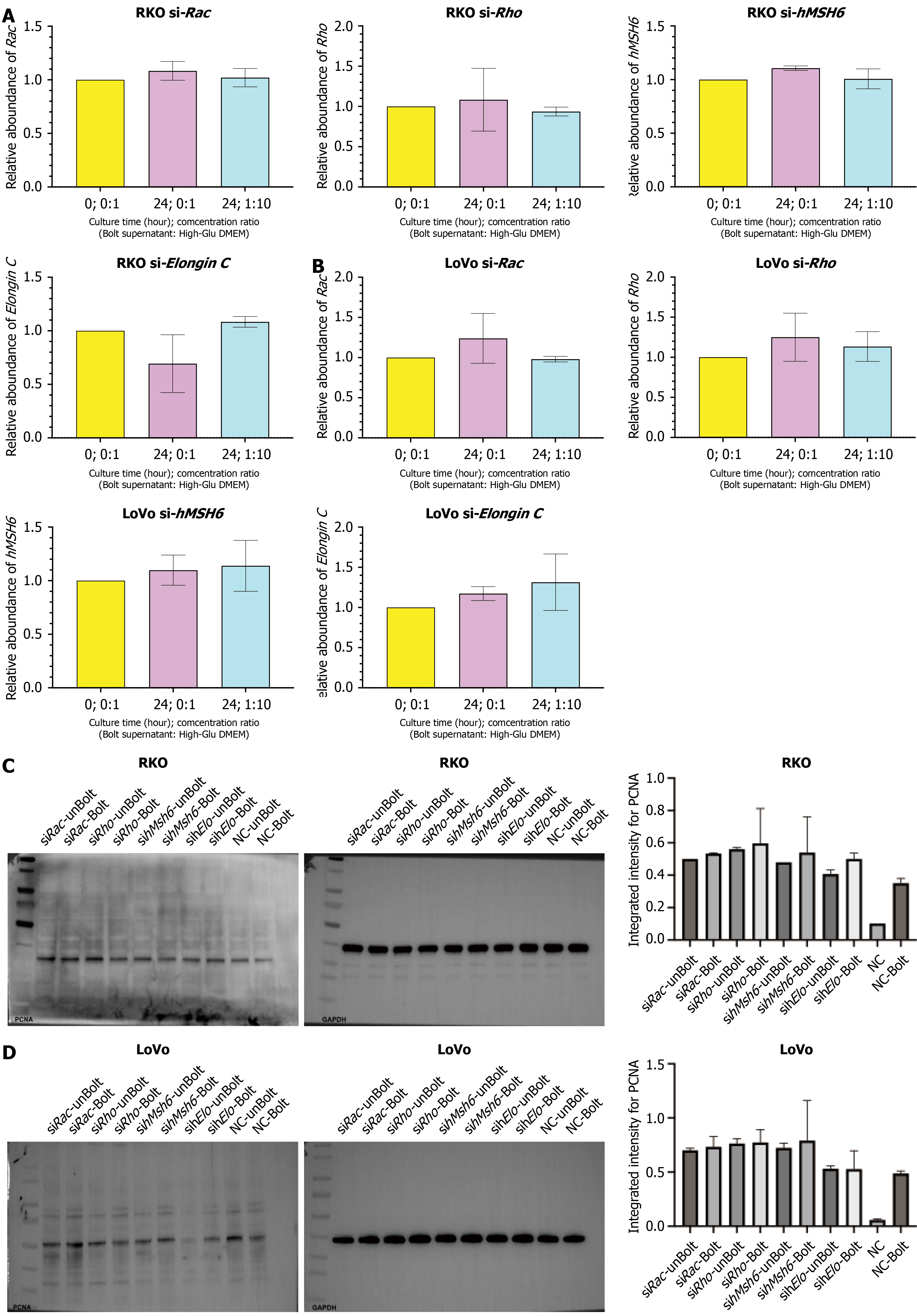
Figure 8 Silencing of Rac, Rho, hMSH6, and Elongin C via siRNA and assessment of the silencing effect on the proliferation marker proliferating cell nuclear antigen in RKO and LoVo colorectal cancer cells.
A: Quantitative real-time polymerase chain reaction quantitative analysis of Rac, Rho, hMSH6, and Elongin C expression levels in RKO cells. The vertical axis represents the relative abundance of these transcripts in colorectal cancer cells, and the horizontal axis represents different intervention conditions (no Bolteae supernatant, or a 1:10 ratio of Bolteae supernatant to culture medium); B: Quantitative real-time polymerase chain reaction quantitative analysis of Rac, Rho, hMSH6, and Elongin C expression levels in LoVo cells; C: Western blot detection of the proliferation marker proliferating cell nuclear antigen (PCNA) in RKO cells transfected with siRNA. The upper panel shows the immunoblot bands, and the lower panel presents the corresponding densitometric quantification. The vertical axis represents the integrated intensity of PCNA, while the horizontal axis indicates different intervention conditions (no Bolteae supernatant, or a 1:10 ratio of Bolteae supernatant to culture medium); D: Western blot detection of the proliferation marker PCNA in LoVo cells transfected with siRNA. PCNA: Proliferating cell nuclear antigen.
- Citation: Lai Y, Xu JH, Chen YJ, Luo H, Lin Y, Yu YF, Chen QK. Enterocloster bolteae drives hyperglycemia-associated colorectal cancer cell proliferation via Rac/Rho-MSH6-Elongin C signaling pathway. World J Gastrointest Oncol 2026; 18(10): 119879
- URL: https://www.wjgnet.com/1948-5204/full/v18/i10/119879.htm
- DOI: https://dx.doi.org/10.4251/wjgo.119879