
















©The Author(s) 2026.
World J Gastrointest Oncol. Jan 15, 2026; 18(1): 114648
Published online Jan 15, 2026. doi: 10.4251/wjgo.v18.i1.114648
Published online Jan 15, 2026. doi: 10.4251/wjgo.v18.i1.114648
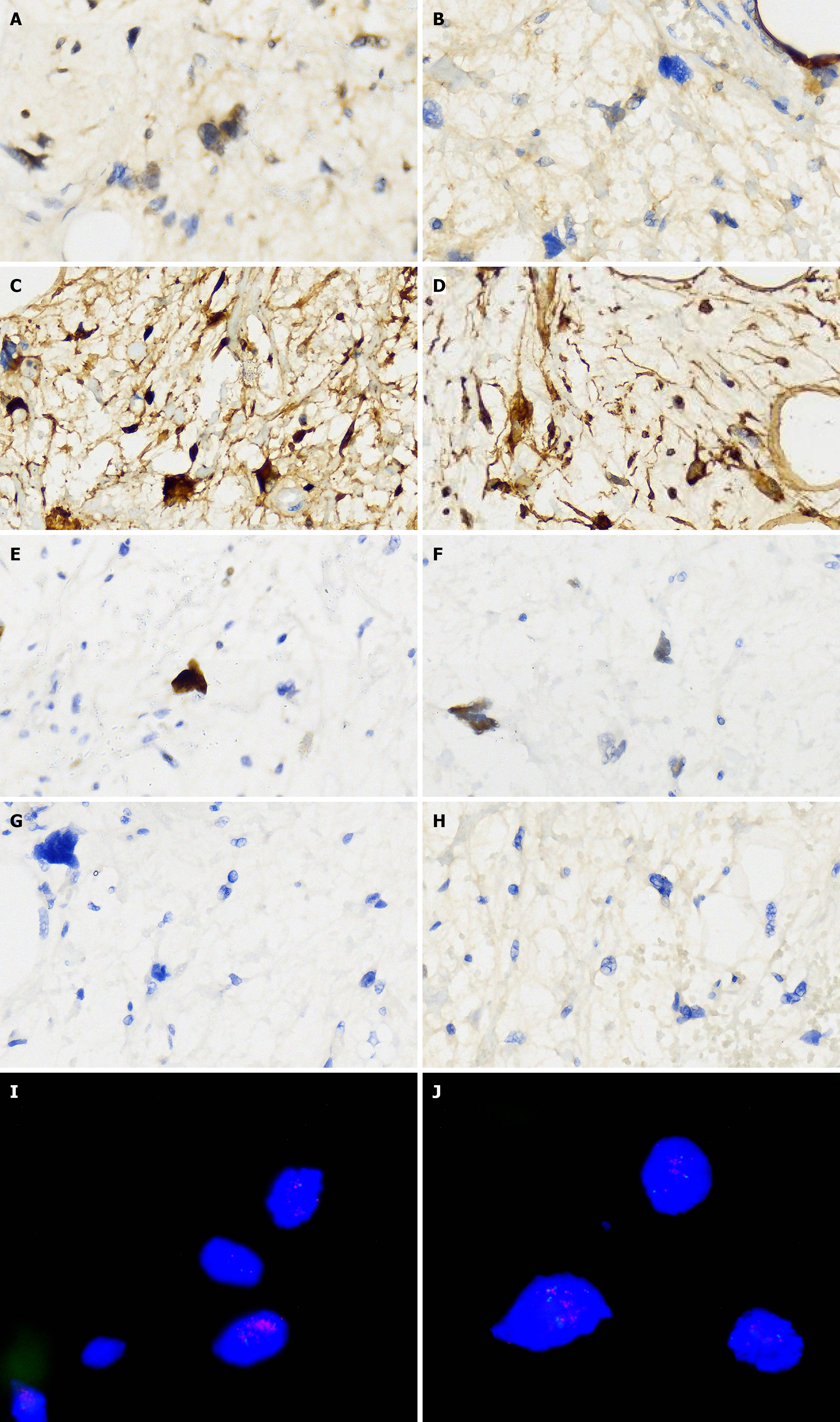
Figure 4 Pathological examination results.
Histopathological examination showed hematoxylin and eosin stains (× 40). Microscopic examination of the mesenteric mass showing variably sized lipoblasts with vacuolated cytoplasm, central hyperchromatic nuclei and nuclear scalloping. Histopathologic features of well-differentiated liposarcoma. A-F: Immunohistochemical stain is positive for the neoplastic cells. Positive: MDM2 (A), S-100 (B), P16 (C), Vim (D), Ki-67 (3%; E), and p53 (F); G and H: Immunohistochemical stain is negative [CK-pan (G) and EMA (H)]; I and J: Fluorescence in situ hybridization showed that MDM2 gene amplification is further confirmed (red signals represent MDM2; green signals represent chromosome 12 centromeres; magnification: × 400).
- Citation: Tian Y, Liu GQ, Li CF, Tian QM, Qiao S. Colossal well-differentiated liposarcoma of the small bowel mesentery: A case report. World J Gastrointest Oncol 2026; 18(1): 114648
- URL: https://www.wjgnet.com/1948-5204/full/v18/i1/114648.htm
- DOI: https://dx.doi.org/10.4251/wjgo.v18.i1.114648